

| 经过测试的应用 | |
| WB | WB-1/500 - 1/2000 |
| FCM | FCM-1/200 - 1/400 |
| IHC | IHC-1/200 - 1/1000 |
| IF/ICC | IF/ICC-1/200 - 1/1000 |
| 反应物种 | Human,Mouse,Rat,Monkey |
| SwissProt ID | Q5VT66 |
| 抗体类型 | Primary antibody |
| 克隆性 | Monoclonal |
| 宿主 | Mouse IgG2a |
| 基因名 | MTARC1 |
| 分子量 | 37kDa |
| 形式 | Liquid |
| 存储缓冲液 | Purified antibody in PBS with 0.05% sodium azide |
| 储存 | Store at 4°C short term. Aliquot and store at -20°C long term. Avoid freeze/thaw cycles. |

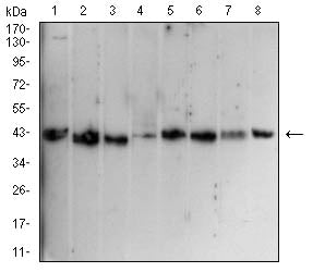
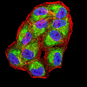
Western blot analysis using MTARC1 mouse mAb against Hela (1), HepG2 (2), HEK293 (3),MOLT4 (4),K562 (5),HEK293-6e (6),Cos-7 (7),and NIH/3T3 (8) cell lysate. Immunofluorescence analysis of Hela cells using MTARC1 mouse mAb (green). Blue: DRAQ5 fluorescent DNA dye. Red: Actin filaments have been labeled with Alexa Fluor- 555 phalloidin. Secondary antibody from Fisher
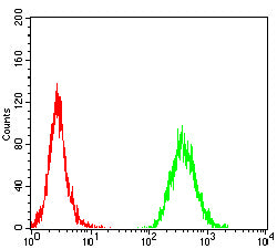
 Flow cytometric analysis of MOLT4 cells using MTARC1 mouse mAb (green) and negative control (red).
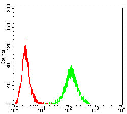
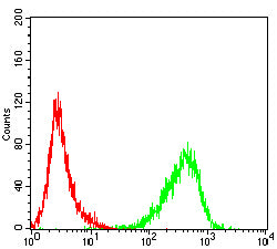
 Flow cytometric analysis of HL-60 cells using MTARC1 mouse mAb (green) and negative control (red).
 Flow cytometric analysis of Jukrat cells using MTARC1 mouse mAb (green) and negative control (red).
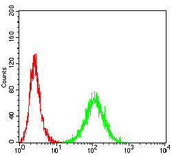
 Flow cytometric analysis of K562 cells using MTARC1 mouse mAb (green) and negative control (red).
 Immunohistochemical analysis of paraffin-embedded human bladder cancer tissues using MTARC1 mouse mAb with DAB staining.
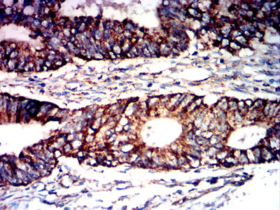
 Immunohistochemical analysis of paraffin-embedded human rectal cancer tissues using MTARC1 mouse mAb with DAB staining.
×
