
| 经过测试的应用 | |
| WB | WB-1:1,000-1:2,000 |
| IF-Cell | IF-Cell-1:100 |
| 反应物种 | Human |
| SwissProt ID | Q16552 |
| 抗体类型 | monoclonal Antibody |
| 阳性对照 | THP-1 cell lysate, HeLa cell lysate, K-562 cell lysate, Jurkat cell lysate, Raji cell lysate, HL-60 cell lysate, HeLa. |
浓度 | 1ug/ul |
| 基因名 | IL17A |
| 分子量 | 96kDa |
| 形式 | Liquid |
| 存储缓冲液 | Purified antibody in PBS with 0.05% sodium azide and 50% glycerol. |
| 储存 | Store at 4°C short term. Aliquot and store at -20°C long term. Avoid freeze/thaw cycles. |

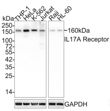
Western blot analysis of IL17A Receptor on different lysates with Rabbit anti-IL17A Receptor antibody at 1/1,000 dilution.
Lane 1: THP-1 cell lysate
Lane 2: HeLa cell lysate
Lane 3: K-562 cell lysate
Lane 4: Jurkat cell lysate
Lane 5: Raji cell lysate
Lane 6: HL-60 cell lysate
Lysates/proteins at 25 µg/Lane.
Predicted band size: 96 kDa
Observed band size: 160 kDa
Exposure time: 30 seconds;
4-20% SDS-PAGE gel.
Proteins were transferred to a PVDF membrane and blocked with 5% NFDM/TBST for 1 hour at room temperature. The primary antibody at 1/1,000 dilution was used in 5% NFDM/TBST at 4℃ overnight. Goat Anti-Rabbit IgG - HRP Secondary Antibody at 1/50,000 dilution was used for 1 hour at room temperature.

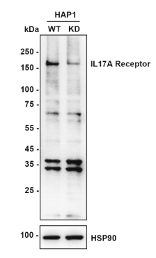
Western blot analysis of IL17A Receptor on different lysates with Rabbit anti-IL17A Receptor antibody at 1/2,000 dilution.
Lane 1: HAP1-parental cell lysate
Lane 2: HAP1-IL17A Receptor KD cell lysate
Lysates/proteins at 10 µg/Lane.
Predicted band size: 96 kDa
Observed band size: 160 kDa
Exposure time: 180 seconds; ECL: K1801;
4-20% SDS-PAGE gel.
Proteins were transferred to a PVDF membrane and blocked with 5% NFDM/TBST for 1 hour at room temperature. The primary antibody at 1/2,000 dilution was used in K1803 at 4℃ overnight. Goat Anti-Rabbit IgG - HRP Secondary Antibody at 1/50,000 dilution was used for 1 hour at room temperature.

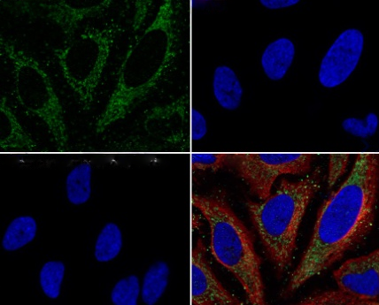
Immunocytochemistry analysis of HeLa cells labeling IL17A Receptor with Rabbit anti-IL17A Receptor antibody at 1/100 dilution.
Cells were fixed in 4% paraformaldehyde for 20 minutes at room temperature, permeabilized with 0.1% Triton X-100 in PBS for 5 minutes at room temperature, then blocked with 1% BSA in 10% negative goat serum for 1 hour at room temperature. Cells were then incubated with Rabbit anti-IL17A Receptor antibody at 1/100 dilution in 1% BSA in PBST overnight at 4 ℃. Goat Anti-Rabbit IgG H&L was used as the secondary antibody at 1/1,000 dilution. PBS instead of the primary antibody was used as the secondary antibody only control. Nuclear DNA was labelled in blue with DAPI.
Beta tubulin was stained at 1/100 dilution overnight at +4℃. Goat Anti-Mouse IgG H&L was used as the secondary antibody at 1/1,000 dilution.
×
