
| WB | 1:2,000 | Human, Mouse, Rat |
| IHC-P | 1:200 | Human, Mouse, Rat |
| IF-Cell | 1:100 | Human, Mouse, Rat |
| FC | 1:1,000 | Human, Mouse, Rat |
| IF-Tissue | 1:200 | Human, Mouse, Rat |
GCH1 Recombinant Rabbit Monoclonal Antibody [PSH08-00]
Recombinant Rabbit monoclonal Antibody
Recombinant protein within human GCH1 aa 1-250.
Human, Mouse, Rat
WB, IHC-P, IF-Cell, FC, IF-Tissue
Predicted band size: 28 kDa
SH-SY5Y cell lysate, HEK-293 cell lysate, Neuro-2a cell lysate, PC-12 cell lysate, Human liver tissue lysate, Mouse liver tissue lysate, Rat liver tissue lysate, human pancreas tissue, mouse pancreas tissue, rat pancreas tissue, Neuro-2a, PC-12.
unconjugated
PSH08-00

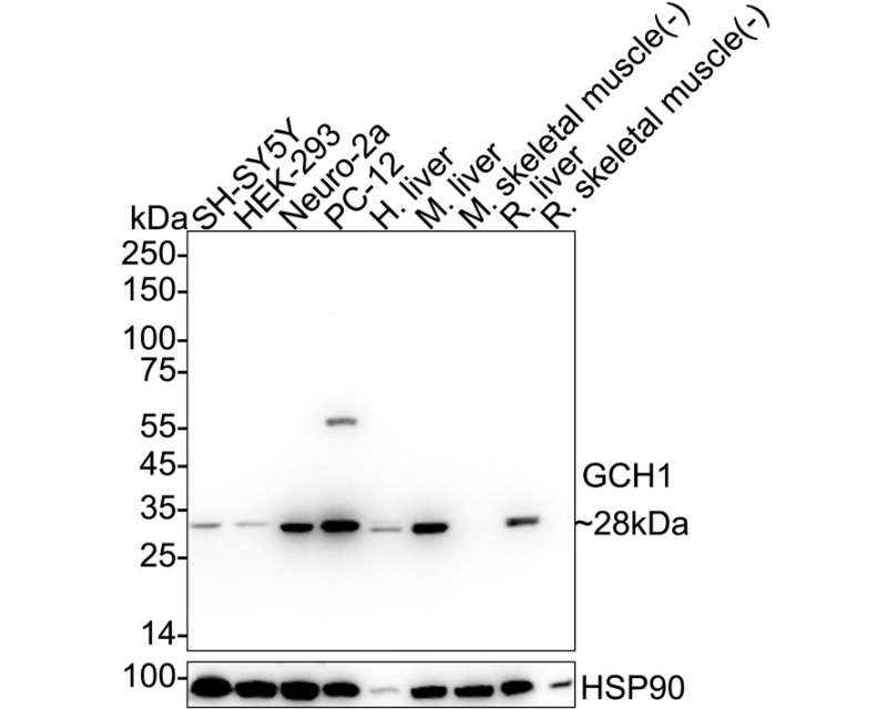
Western blot analysis of GCH1 on different lysates with Rabbit anti-GCH1 antibody at 1/2,000 dilution.
Lane 1: SH-SY5Y cell lysate
Lane 2: HEK-293 cell lysate
Lane 3: Neuro-2a cell lysate
Lane 4: PC-12 cell lysate
Lane 5: Human liver tissue lysate
Lane 6: Mouse liver tissue lysate
Lane 7: Mouse skeletal muscle tissue lysate (negative)
Lane 8: Rat liver tissue lysate
Lane 9: Rat skeletal muscle tissue lysate (negative)
Lysates/proteins at 20 µg/Lane.
Predicted band size: 28 kDa
Observed band size: 28 kDa
Exposure time: 25 seconds; ECL: K1801;
4-20% SDS-PAGE gel.
Proteins were transferred to a PVDF membrane and blocked with 5% NFDM/TBST for 1 hour at room temperature. The primary antibody at 1/2,000 dilution was used in 5% NFDM/TBST at 4℃ overnight. Goat Anti-Rabbit IgG - HRP Secondary Antibody at 1/50,000 dilution was used for 1 hour at room temperature.

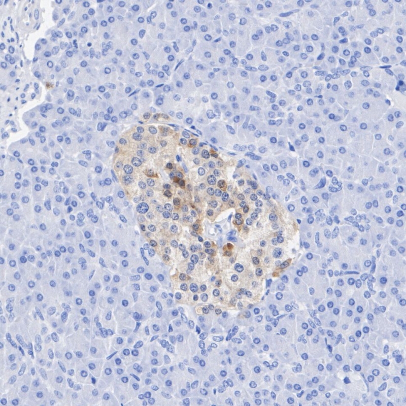
Immunohistochemical analysis of paraffin-embedded human pancreas tissue with Rabbit anti-GCH1 antibody at 1/200 dilution.
The section was pre-treated using heat mediated antigen retrieval with sodium citrate buffer (pH 6.0) for 2 minutes. The tissues were blocked in 1% BSA for 20 minutes at room temperature, washed with ddH2O and PBS, and then probed with the primary antibody at 1/200 dilution for 1 hour at room temperature. The detection was performed using an HRP conjugated compact polymer system. DAB was used as the chromogen. Tissues were counterstained with hematoxylin and mounted with DPX.

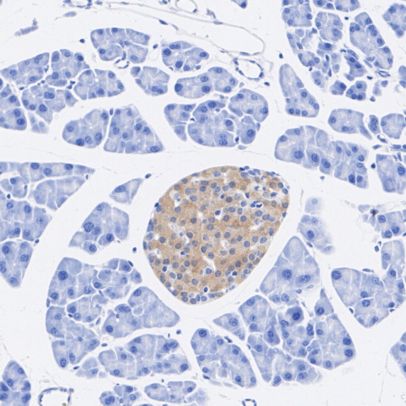
Immunohistochemical analysis of paraffin-embedded mouse pancreas tissue with Rabbit anti-GCH1 antibody at 1/200 dilution.
The section was pre-treated using heat mediated antigen retrieval with sodium citrate buffer (pH 6.0) for 2 minutes. The tissues were blocked in 1% BSA for 20 minutes at room temperature, washed with ddH2O and PBS, and then probed with the primary antibody at 1/200 dilution for 1 hour at room temperature. The detection was performed using an HRP conjugated compact polymer system. DAB was used as the chromogen. Tissues were counterstained with hematoxylin and mounted with DPX.

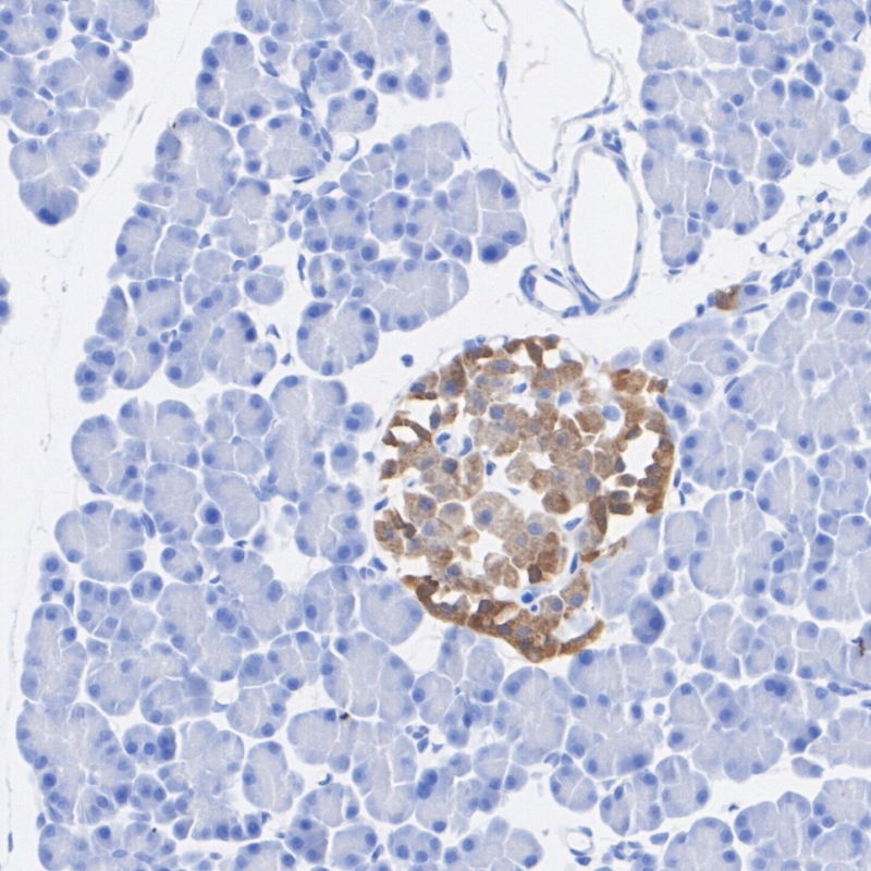
Immunohistochemical analysis of paraffin-embedded rat pancreas tissue with Rabbit anti-GCH1 antibody at 1/200 dilution.
The section was pre-treated using heat mediated antigen retrieval with sodium citrate buffer (pH 6.0) for 2 minutes. The tissues were blocked in 1% BSA for 20 minutes at room temperature, washed with ddH2O and PBS, and then probed with the primary antibody at 1/200 dilution for 1 hour at room temperature. The detection was performed using an HRP conjugated compact polymer system. DAB was used as the chromogen. Tissues were counterstained with hematoxylin and mounted with DPX.

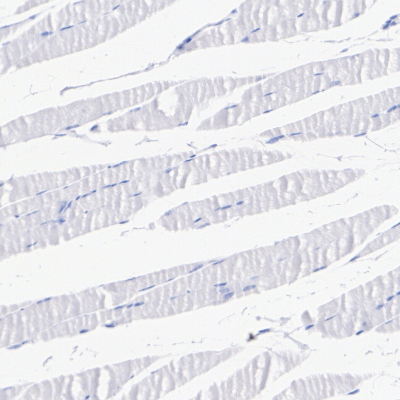
Immunohistochemical analysis of paraffin-embedded human skeletal muscle tissue (negative) with Rabbit anti-GCH1 antibody at 1/200 dilution.
The section was pre-treated using heat mediated antigen retrieval with sodium citrate buffer (pH 6.0) for 2 minutes. The tissues were blocked in 1% BSA for 20 minutes at room temperature, washed with ddH2O and PBS, and then probed with the primary antibody at 1/200 dilution for 1 hour at room temperature. The detection was performed using an HRP conjugated compact polymer system. DAB was used as the chromogen. Tissues were counterstained with hematoxylin and mounted with DPX.

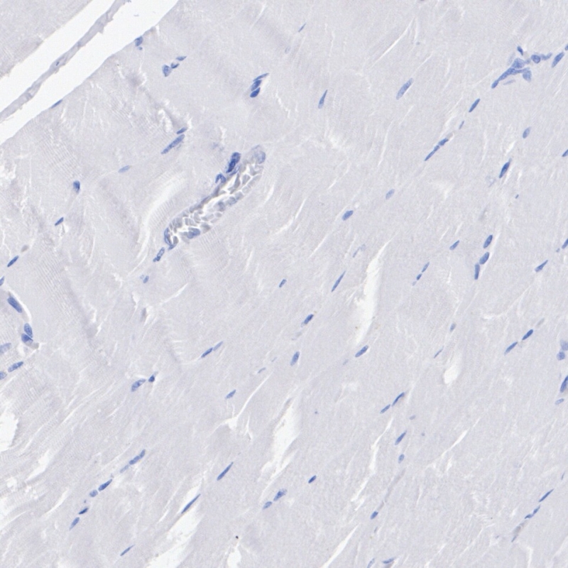
Immunohistochemical analysis of paraffin-embedded rat skeletal muscle tissue (negative) with Rabbit anti-GCH1 antibody at 1/200 dilution.
The section was pre-treated using heat mediated antigen retrieval with sodium citrate buffer (pH 6.0) for 2 minutes. The tissues were blocked in 1% BSA for 20 minutes at room temperature, washed with ddH2O and PBS, and then probed with the primary antibody at 1/200 dilution for 1 hour at room temperature. The detection was performed using an HRP conjugated compact polymer system. DAB was used as the chromogen. Tissues were counterstained with hematoxylin and mounted with DPX.

Immunofluorescence analysis of paraffin-embedded mouse pancreas tissue labeling GCH1 with Rabbit anti-GCH1 antibody at 1/200 dilution.
The section was pre-treated using heat mediated antigen retrieval with sodium citrate buffer (pH 6.0) for 2 minutes. The tissues were blocked in 10% negative goat serum for 1 hour at room temperature, washed with PBS, and then probed with the primary antibody ( green) at 1/200 dilution overnight at 4 ℃, washed with PBS. Goat Anti-Rabbit IgG H&L (iFluor™ 488, ) was used as the secondary antibody at 1/1,000 dilution. Nuclei were counterstained with DAPI (blue).

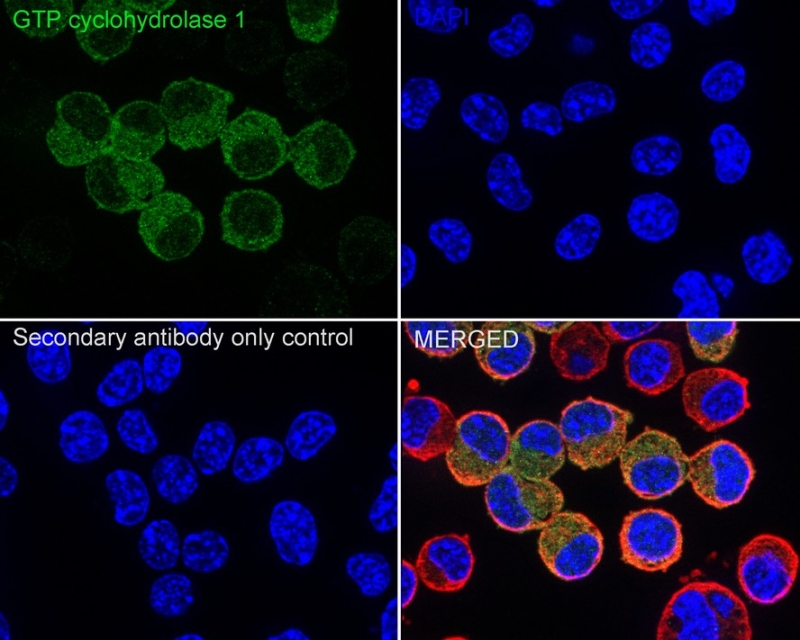
Immunocytochemistry analysis of Neuro-2a cells labeling GCH1 with Rabbit anti-GCH1 antibody at 1/100 dilution.
Cells were fixed in 4% paraformaldehyde for 15 minutes at room temperature, permeabilized with 0.1% Triton X-100 in PBS for 15 minutes at room temperature, then blocked with 1% BSA in 10% negative goat serum for 1 hour at room temperature. Cells were then incubated with Rabbit anti-GCH1 antibody at 1/100 dilution in 1% BSA in PBST overnight at 4 ℃. Goat Anti-Rabbit IgG H&L (iFluor™ 488, ) was used as the secondary antibody at 1/1,000 dilution. PBS instead of the primary antibody was used as the secondary antibody only control. Nuclear DNA was labelled in blue with DAPI.
Beta tubulin (red) was stained at 1/100 dilution overnight at +4℃. Goat Anti-Mouse IgG H&L (iFluor™ 594) was used as the secondary antibody at 1/1,000 dilution.

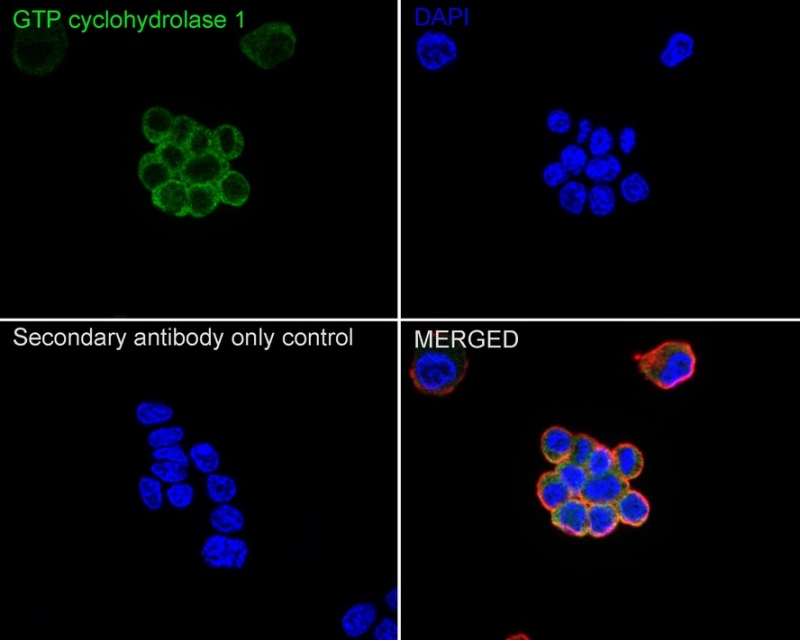
Immunocytochemistry analysis of PC-12 cells labeling GCH1 with Rabbit anti-GCH1 antibody at 1/100 dilution.
Cells were fixed in 4% paraformaldehyde for 15 minutes at room temperature, permeabilized with 0.1% Triton X-100 in PBS for 15 minutes at room temperature, then blocked with 1% BSA in 10% negative goat serum for 1 hour at room temperature. Cells were then incubated with Rabbit anti-GCH1 antibody at 1/100 dilution in 1% BSA in PBST overnight at 4 ℃. Goat Anti-Rabbit IgG H&L (iFluor™ 488) was used as the secondary antibody at 1/1,000 dilution. PBS instead of the primary antibody was used as the secondary antibody only control. Nuclear DNA was labelled in blue with DAPI.
Beta tubulin ( red) was stained at 1/100 dilution overnight at +4℃. Goat Anti-Mouse IgG H&L (iFluor™ 594) was used as the secondary antibody at 1/1,000 dilution.

Flow cytometric analysis of Neuro-2a cells labeling GCH1.
Cells were fixed and permeabilized. Then stained with the primary antibody ( 1μg/mL) (red) compared with Rabbit IgG Isotype Control (green). After incubation of the primary antibody at +4℃ for an hour, the cells were stained with a iFluor™ 488 conjugate-Goat anti-Rabbit IgG Secondary antibody at 1/1,000 dilution for 30 minutes at +4℃. Unlabelled sample was used as a control (cells without incubation with primary antibody; black).
×

