
| 经过测试的应用 | |
| WB | WB-1-2000 |
| IHC-P | IHC-P 1-2000 |
| IF | IF-1-100 |
| 反应物种 | Human, Mouse, Rat |
| SwissProt ID | PSH05-68 |
| 抗体类型 | Monoclonal antibody |
| 克隆性 | Monoclonal |
| 宿主 | Rabbit |
| 基因名 | MUSK |
| 分子量 | 97kDa |
| 背景文献 | 1. Rodolico C et al. MuSK-Associated Myasthenia Gravis: Clinical Features and Management. 2. Fish LA et al. Multiple MuSK signaling pathways and the aging neuromuscular junction. |
| 形式 | Liquid |
| 存储缓冲液 | PBS (pH7.4), 0.1% BSA, 40% Glycerol. Preservative: 0.05% Sodium Azide. |
| 储存 | Store at 4°C short term. Aliquot and store at -20°C long term. Avoid freeze/thaw cycles. |

Immunohistochemical analysis of paraffin-embedded human skeletal muscle tissue with Rabbit anti-MUSK antibody at 1/2,000 dilution.
The section was pre-treated using heat mediated antigen retrieval with Tris-EDTA buffer (pH 9.0) for 20 minutes. The tissues were blocked in 1% BSA for 20 minutes at room temperature, washed with ddH2O and PBS, and then probed with the primary antibody at 1/2,000 dilution for 1 hour at room temperature. The detection was performed using an HRP conjugated compact polymer system. DAB was used as the chromogen. Tissues were counterstained with hematoxylin and mounted with DPX.
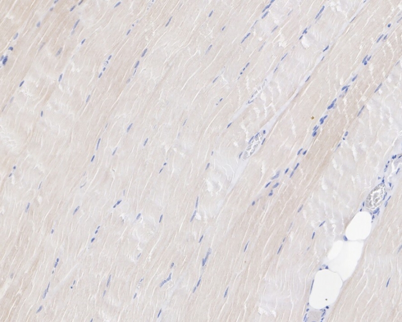

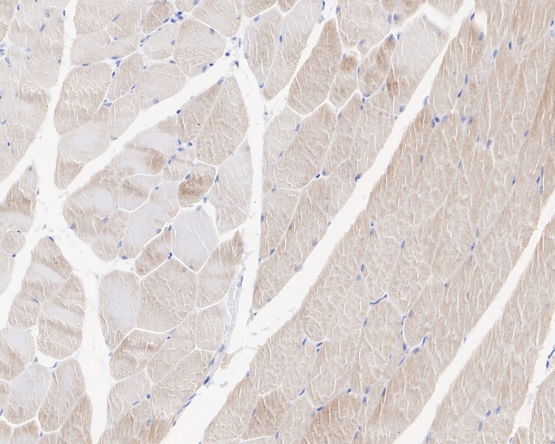
Immunohistochemical analysis of paraffin-embedded mouse skeletal muscle tissue with Rabbit anti-MUSK antibody at 1/2,000 dilution.
The section was pre-treated using heat mediated antigen retrieval with Tris-EDTA buffer (pH 9.0) for 20 minutes. The tissues were blocked in 1% BSA for 20 minutes at room temperature, washed with ddH2O and PBS, and then probed with the primary antibody at 1/2,000 dilution for 1 hour at room temperature. The detection was performed using an HRP conjugated compact polymer system. DAB was used as the chromogen. Tissues were counterstained with hematoxylin and mounted with DPX.

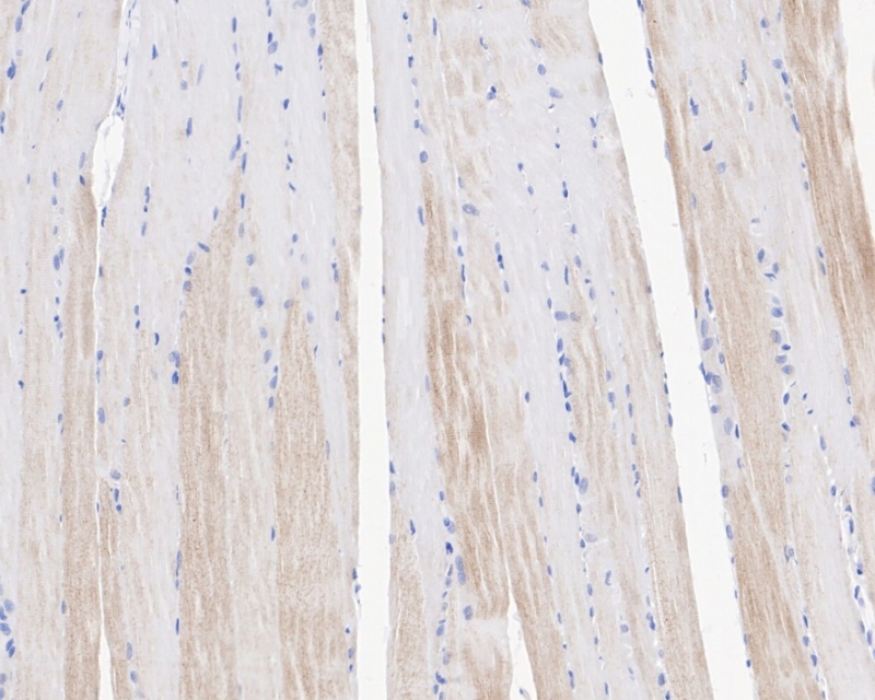
Immunohistochemical analysis of paraffin-embedded rat skeletal muscle tissue with Rabbit anti-MUSK antibody at 1/2,000 dilution.
The section was pre-treated using heat mediated antigen retrieval with Tris-EDTA buffer (pH 9.0) for 20 minutes. The tissues were blocked in 1% BSA for 20 minutes at room temperature, washed with ddH2O and PBS, and then probed with the primary antibody at 1/2,000 dilution for 1 hour at room temperature. The detection was performed using an HRP conjugated compact polymer system. DAB was used as the chromogen. Tissues were counterstained with hematoxylin and mounted with DPX.

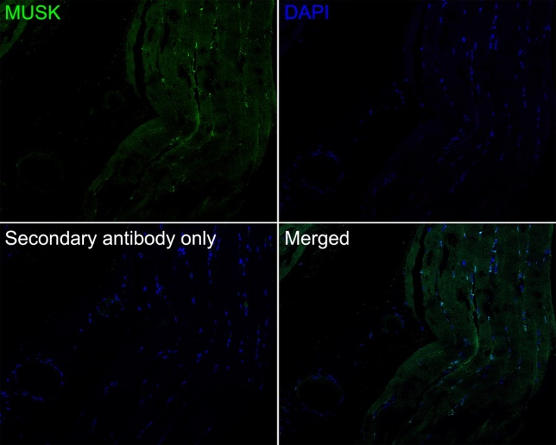
Immunofluorescence analysis of paraffin-embedded human skeletal muscle tissue labeling MUSK with Rabbit anti-MUSK antibody at 1/100 dilution.
The section was pre-treated using heat mediated antigen retrieval with Tris-EDTA buffer (pH 9.0) for 20 minutes. The tissues were blocked in 10% negative goat serum for 1 hour at room temperature, washed with PBS, and then probed with the primary antibody at 1/100 dilution overnight at 4 ℃, washed with PBS. Goat Anti-Rabbit IgG H&L (iFluor™ 488,) was used as the secondary antibody at 1/1,000 dilution. Nuclei were counterstained with DAPI (blue).
×
