
| WB | 1:500-2000 | mouse, rat |
| FCM | 1:50-200 | mouse, rat |
| IHC | 1:50-400 | mouse, rat |
12 months from date of receipt,-20℃ as supplied.

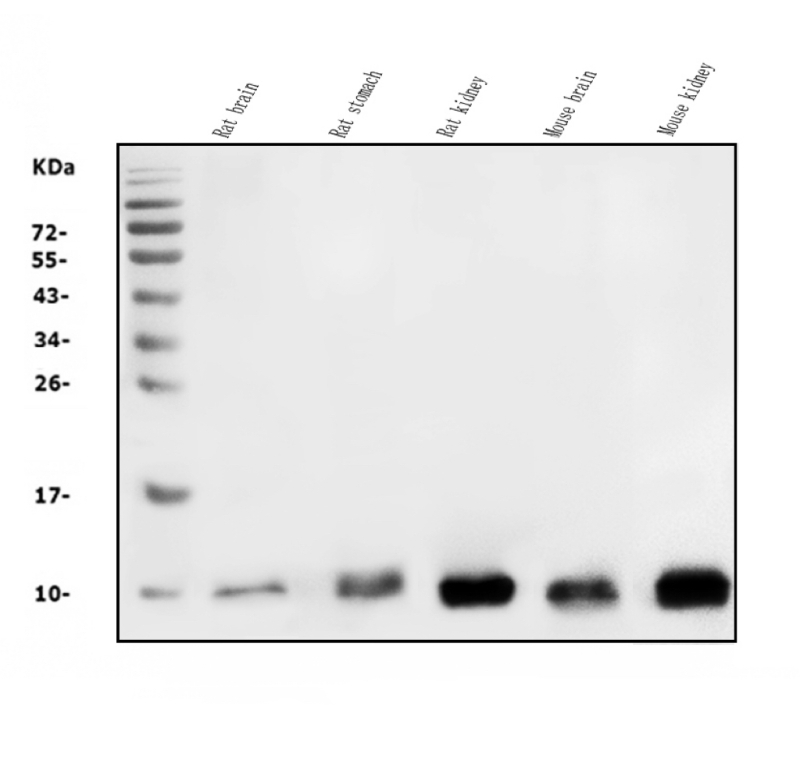
Western blot analysis of COX8A using anti-COX8A antibody . The
sample well of each lane was loaded with 30 ug of sample under reducing
conditions.
Lane 1: Rat brain tissue lysates,
Lane 2: Rat stomach tissue lysates,
Lane 3: Rat kidney tissue lysates,
Lane 4: Mouse brain tissue lysates,
Lane 5: Mouse kidney tissue lysates.
After
electrophoresis, proteins were transferred to a membrane. Then the
membrane was incubated with rabbit anti-COX8A antigen affinity purified
polyclonal antibody at a dilution of 1:1000 and probed with a
goat anti-rabbit IgG-HRP secondary antibody . The
signal is developed using ECL Plus Western Blotting Substrate. A specific band was detected for COX8A at approximately 8-10
kDa. The expected band size for COX8A is at 8 kDa.

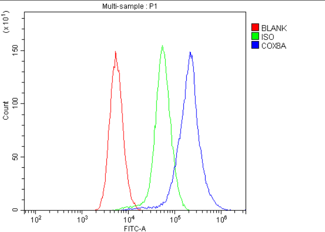
Flow Cytometry analysis of mouse spleen tissue using anti-COX8A antibody .
Overlay
histogram showing mouse spleen tissue stained with (Blue
line). To facilitate intracellular staining, tissue was fixed with 4%
paraformaldehyde and permeabilized with permeabilization buffer. The
tissue was blocked with 10% normal goat serum. And then incubated with
rabbit anti-COX8A Antibody at 1:100 dilution for 30 min at
20°C. DyLight®488 conjugated goat anti-rabbit IgG was used as
secondary antibody at 1:100 dilution for 30 minutes at 20°C. Isotype
control antibody (Green line) was rabbit IgG at 1:100 dilution used
under the same conditions. Unlabelled sample without incubation with
primary antibody and secondary antibody (Red line) was used as a blank
control.

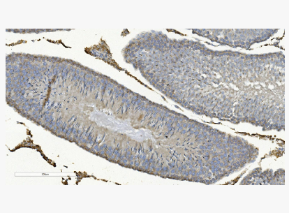
IHC analysis of COX8A using anti-COX8A antibody .
COX8A
was detected in a paraffin-embedded section of rat testis tissue.
Biotinylated goat anti-rabbit IgG was used as secondary antibody. The
tissue section was incubated with rabbit anti-COX8A Antibody at a dilution of 1:200 and developed using Strepavidin-Biotin-Complex
(SABC) with DAB as the chromogen.
×
