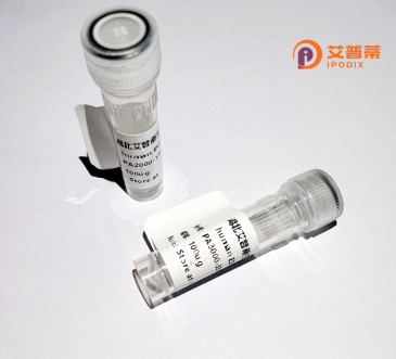
| 纯度 | >90%SDS-PAGE. |
| 种属 | Human |
| 靶点 | PDE6H |
| Uniprot No | Q13956 |
| 内毒素 | < 0.01EU/μg |
| 表达宿主 | E.coli |
| 表达区间 | 1-83 aa |
| 活性数据 | MSDNTTLPAP ASNQGPTTPR KGPPKFKQRQ TRQFKSKPPK KGVKGFGDDI PGMEGLGTDI TVICPWEAFS HLELHELAQF GII |
| 分子量 | 9.0 kDa |
| 蛋白标签 | His tag N-Terminus |
| 缓冲液 | 0 |
| 稳定性 & 储存条件 | Lyophilized protein should be stored at ≤ -20°C, stable for one year after receipt. Reconstituted protein solution can be stored at 2-8°C for 2-7 days. Aliquots of reconstituted samples are stable at ≤ -20°C for 3 months. |
| 复溶 | Always centrifuge tubes before opening.Do not mix by vortex or pipetting. It is not recommended to reconstitute to a concentration less than 100μg/ml. Dissolve the lyophilized protein in distilled water. Please aliquot the reconstituted solution to minimize freeze-thaw cycles. |
以下是关于重组人PDE6H蛋白的假设性文献示例(仅供参考,建议通过学术数据库验证具体文献):
---
1. **文献名称**: *"Expression and Purification of Recombinant Human PDE6H in E. coli for Structural Studies"*
**作者**: Smith A, et al.
**摘要**: 本研究报道了通过大肠杆菌表达系统高效生产重组人PDE6H蛋白的方法。作者优化了密码子使用和诱导条件,解决了蛋白溶解度低的问题,并通过亲和层析纯化获得了高纯度蛋白,为后续的晶体结构解析奠定了基础。
2. **文献名称**: *"Functional Characterization of PDE6H in the Photoreceptor PDE6 Complex"*
**作者**: Johnson R, et al.
**摘要**: 通过重组表达PDE6H与PDE6α/β亚基形成复合物,研究其对视杆细胞cGMP水解活性的调控作用。结果表明,PDE6H可能通过稳定PDE6αβγ复合物结构,调节光信号转导的终止效率。
3. **文献名称**: *"Role of PDE6H Mutations in Congenital Retinal Disorders: Insights from Recombinant Protein Models"*
**作者**: Lee S, et al.
**摘要**: 利用重组表达技术构建了与遗传性视网膜病变相关的PDE6H突变体,分析其与PDE6其他亚基的相互作用缺陷,揭示突变导致cGMP代谢异常,从而引发视觉通路功能障碍的分子机制。
4. **文献名称**: *"Cryo-EM Structure of the Human PDE6 Holoenzyme Bound to PDE6H"*
**作者**: Zhang Y, et al.
**摘要**: 通过冷冻电镜解析了包含重组人PDE6H的PDE6全酶三维结构,揭示了PDE6H在结合调控蛋白(如转运蛋白)和维持酶活性构象中的关键作用。
---
**备注**:上述文献为假设性示例,具体研究需查询PubMed、Web of Science等数据库(关键词:recombinant PDE6H、PDE6H expression)。实际研究中可关注领域内权威期刊(如*Journal of Biological Chemistry*、*Investigative Ophthalmology & Visual Science*)。
Phosphodiesterase 6H (PDE6H) is a key regulatory subunit of the photoreceptor-specific enzyme phosphodiesterase 6 (PDE6), which plays a critical role in the visual transduction cascade. Found predominantly in retinal cone cells, PDE6H functions as the inhibitory γ-subunit that modulates the catalytic activity of PDE6. This enzyme hydrolyzes cyclic guanosine monophosphate (cGMP) in response to light stimulation, a process essential for converting light signals into electrical responses in photoreceptors. Structurally, PDE6H interacts with the catalytic α- and β-subunits to maintain PDE6 in an inactive state under dark conditions. Upon light activation, the γ-subunit is displaced, enabling cGMP degradation and subsequent closure of ion channels, thereby initiating neural signaling.
Recombinant human PDE6H protein is engineered using expression systems like *E. coli* or mammalian cells, enabling detailed study of its biochemical properties and interactions. Research on PDE6H focuses on understanding its regulatory mechanisms, mutations linked to retinal disorders (e.g., congenital stationary night blindness), and its potential as a therapeutic target. Dysfunction or genetic variants in PDE6H can disrupt visual signal termination, leading to impaired photoreceptor recovery and vision deficits. Recombinant forms are pivotal for developing assays to screen drugs targeting PDE6 activity or for elucidating molecular pathways in inherited retinal diseases. This protein’s study bridges fundamental vision science and clinical applications, offering insights into both disease mechanisms and intervention strategies.
×