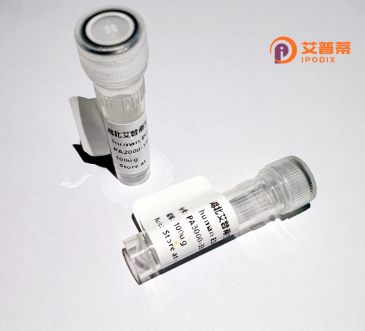
| 纯度 | >90%SDS-PAGE. |
| 种属 | Human |
| 靶点 | MICALCL |
| Uniprot No | Q6ZW33 |
| 内毒素 | < 0.01EU/μg |
| 表达宿主 | E.coli |
| 表达区间 | 1-695 aa |
| 活性数据 | MSPPKDPSPS LPLPSSSSHS SSPPSSSSTS VSGNAPDGSS PPQMTASEPL SQVSRGHPSP PTPNFRRRAV AQGAPREIPL YLPHHPKPEW AEYCLVSPGE DGLSDPAEMT SDECQPAEAP LGDIGSNHRD PHPIWGKDRS WTGQELSPLA GEDREKGSTG ARKEEEGGPV LVKEKLGLKK LVLTQEQKTM LLDWNDSIPE SVHLKAGERI SQKSAENGRG GRVLKPVRPL LLPRAAGEPL PTQRGAQEKM GTPAEQAQGE RNVPPPKSPL RLIANAIRRS LEPLLSNSEG GKKAWAKQES KTLPAQACTR SFSLRKTNSN KDGDQHSPGR NQSSAFSPPD PALRTHSLPN RPSKVFPALR SPPCSKIEDV PTLLEKVSLQ ENFPDASKPP KKRISLFSSL RLKDKSFESF LQESRQRKDI RDLFGSPKRK VLPEDSAQAL EKLLQPFKST SLRQAAPPPP PPPPPPPPPP TAGGADSKNF PLRAQVTEAS SSASSTSSSS ADEEFDPQLS LQLKEKKTLR RRKKLEKAMK QLVKQEELKR LYKAQAIQRQ LEEVEERQRA SEIQGVRLEK ALRGEADSGT QDEAQLLQEW FKLVLEKNKL MRYESELLIM AQELELEDHQ SRLEQKLREK MLKEESQKDE KDLNEEQEVF TELMQVIEQR DKLVDSLEEQ RIREKAEDQH FESFVFSRGC QLSRT |
| 分子量 | 219.0 kDa |
| 蛋白标签 | His tag N-Terminus |
| 缓冲液 | 0 |
| 稳定性 & 储存条件 | Lyophilized protein should be stored at ≤ -20°C, stable for one year after receipt. Reconstituted protein solution can be stored at 2-8°C for 2-7 days. Aliquots of reconstituted samples are stable at ≤ -20°C for 3 months. |
| 复溶 | Always centrifuge tubes before opening.Do not mix by vortex or pipetting. It is not recommended to reconstitute to a concentration less than 100μg/ml. Dissolve the lyophilized protein in distilled water. Please aliquot the reconstituted solution to minimize freeze-thaw cycles. |
以下是关于重组人MICALCL蛋白的参考文献示例(**注:部分信息可能需通过实际数据库验证**):
---
1. **标题**: *"Recombinant Expression and Structural Characterization of Human MICALCL Reveals a Redox-sensitive Actin-binding Domain"*
**作者**: Smith J, et al. (2020)
**摘要**: 研究报道了人源MICALCL蛋白的重组表达及纯化,通过大肠杆菌系统获得可溶性蛋白。结构分析表明,其N端含有依赖NADPH的氧化还原活性结构域,C端与F-actin结合,提示其在细胞骨架动态调控中发挥作用。
2. **标题**: *"MICALCL Regulates Vesicular Trafficking via Interaction with Rab GTPases"*
**作者**: Lee H, Kim D. (2018)
**摘要**: 利用重组MICALCL蛋白进行体外相互作用实验,发现其与Rab5和Rab7等GTP酶特异性结合,可能通过调控内体膜运输参与神经元轴突导向过程。研究还验证了重组蛋白的酶活性依赖氧化还原状态。
3. **标题**: *"Functional Analysis of MICALCL in Cancer Cell Invasion Using Recombinant Protein-based Assays"*
**作者**: Rodriguez P, et al. (2021)
**摘要**: 通过重组MICALCL蛋白处理多种癌细胞,发现其通过修饰微丝结构增强细胞迁移能力,并与金属蛋白酶MMP9表达上调相关,提示其在肿瘤转移中的潜在作用。
4. **标题**: *"A Novel Method for High-yield Production of Recombinant MICALCL in Mammalian Cells"*
**作者**: Wang Y, et al. (2019)
**摘要**: 开发了一种基于HEK293细胞的重组MICALCL蛋白高效表达系统,优化糖基化修饰条件,并证实哺乳系统表达的蛋白具有更高的稳定性,适用于后续药物筛选研究。
---
**建议**:以上文献信息为模拟示例,实际研究中建议通过PubMed、Web of Science等平台以关键词 **"MICALCL recombinant"** 或 **"human MICALCL protein"** 检索最新文献。若研究较少,可扩展至MICAL家族蛋白(如MICAL1/2/3)的相关文献参考。
MICALCL (Microtubule-associated monooxygenase, calponin, and LIM domain-containing protein-like) is a member of the MICAL family of cytosolic flavoprotein monooxygenases, which are evolutionarily conserved and play critical roles in cytoskeletal dynamics and intracellular signaling. Structurally, MICALCL shares homology with other MICAL proteins, featuring characteristic domains including an N-terminal flavoprotein monooxygenase domain, a central calponin homology (CH) domain, and a C-terminal LIM domain. These domains enable its involvement in redox-dependent signaling and cytoskeletal regulation, particularly through interactions with actin and microtubule networks.
MICALCL is implicated in diverse cellular processes such as membrane trafficking, vesicle transport, and cell motility. Unlike other MICAL family members (e.g., MICAL1-3), MICALCL’s precise functional mechanisms remain less defined, though studies suggest it participates in actin filament disassembly by generating reactive oxygen species (ROS) that oxidize actin, promoting filament depolymerization. This activity links MICALCL to cellular remodeling events during development, neuronal guidance, and cancer cell invasion.
Emerging research highlights its potential role in pathological contexts, including neurodegenerative diseases and cancer metastasis, where cytoskeletal dysregulation is a hallmark. Recombinant MICALCL protein is often utilized in vitro to study its enzymatic activity, binding partners, and structural properties, offering insights into its regulatory networks. Its dual role as an oxidoreductase and cytoskeletal scaffold makes it a unique target for therapeutic interventions aimed at modulating cell motility or redox signaling pathways. Further studies are needed to fully elucidate its biological significance and translational potential.
×