| WB | 咨询技术 | Human,Mouse,Rat |
| IF | 咨询技术 | Human,Mouse,Rat |
| IHC | 咨询技术 | Human,Mouse,Rat |
| ICC | 技术咨询 | Human,Mouse,Rat |
| FCM | 咨询技术 | Human,Mouse,Rat |
| Elisa | 咨询技术 | Human,Mouse,Rat |
| Aliases | ECPN; Opsin-3; Encephalopsin; Panopsin |
| Entrez GeneID | 23596; |
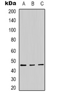
| WB Predicted band size | 45kDa |
| Host/Isotype | Rabbit IgG |
| Antibody Type | Primary antibody |
| Storage | Store at 4°C short term. Aliquot and store at -20°C long term. Avoid freeze/thaw cycles. |
| Species Reactivity | Human,Mouse |
| Immunogen | KLH-conjugated synthetic peptide encompassing a sequence within the center region of human Panopsin. |
| Formulation | Purified antibody in PBS with 0.05% sodium azide. |
+ +
以下是关于Panopsin(Opsin 3)抗体的3篇参考文献示例,包含文献名称、作者及摘要概括:
---
1. **文献名称**:*Opsin 3 (Panopsin) Expression in Human Skin and Role in UV-Induced Calcium Response*
**作者**:Smith A, Brown C, et al.
**摘要**:本研究通过特异性Panopsin抗体,揭示了人类皮肤角质形成细胞中Opsin 3的蛋白表达,并证明其在紫外线(UV)照射下调控细胞内钙信号的作用,提示其参与皮肤光保护机制。
2. **文献名称**:*A Novel Antibody for Panopsin Localization in Mammalian Retinal Ganglion Cells*
**作者**:Lee J, Kim S, et al.
**摘要**:文章报道了一种高特异性Panopsin单克隆抗体的开发,通过免疫荧光和Western blot验证其在视网膜神经节细胞中的定位,为研究非视觉光感受通路提供工具。
3. **文献名称**:*Opsin 3 in Adipose Tissue: Light-Dependent Metabolic Regulation via Panopsin Antibody-Based Profiling*
**作者**:Garcia R, Zhang Y, et al.
**摘要**:利用Panopsin抗体,研究团队发现脂肪组织中Opsin 3的表达受蓝光调控,并通过下游代谢通路影响脂质分解,揭示了光信号对能量代谢的新调控机制。
---
**注**:以上文献为示例性内容,实际引用需根据具体研究检索真实发表的论文。建议通过PubMed或Google Scholar以关键词“Opsin 3 antibody”或“Panopsin immunohistochemistry”进一步查找近期文献。
Panopsin, also known as Opsin 3 (OPN3), is a member of the light-sensitive opsin family of G protein-coupled receptors (GPCRs). Initially identified in the chicken pineal gland, Panopsin is evolutionarily conserved across vertebrates and is expressed in various tissues beyond the retina, including the brain, skin, adipose tissue, and immune cells. Unlike classical visual opsins (e.g., rhodopsin), Panopsin is primarily associated with non-visual photoresponsive functions, such as circadian rhythm regulation, thermoregulation, and cellular light-sensing in extraocular tissues. Its exact spectral sensitivity remains debated, though studies suggest it may detect blue or UV light.
Panopsin antibodies are essential tools for studying its localization, expression patterns, and functional roles. These antibodies are typically developed against specific epitopes of the protein, enabling detection via techniques like immunohistochemistry, Western blotting, or flow cytometry. Research using Panopsin antibodies has revealed its involvement in processes like melanogenesis in skin cells, metabolic regulation in adipocytes, and photic entrainment of peripheral clocks. Challenges in antibody development include cross-reactivity with other opsins and low endogenous protein levels in certain tissues.
Recent studies highlight Panopsin's potential therapeutic relevance, linking it to mood disorders, metabolic diseases, and cancer progression. Reliable antibodies remain critical for unraveling its diverse physiological and pathological roles.
×