
| WB | 1/1000 | Human,Mouse,Rat |
| IF | 咨询技术 | Human,Mouse,Rat |
| IHC | 咨询技术 | Human,Mouse,Rat |
| ICC | 技术咨询 | Human,Mouse,Rat |
| FCM | 咨询技术 | Human,Mouse,Rat |
| Elisa | 咨询技术 | Human,Mouse,Rat |
| Aliases | DDB1- and CUL4-associated factor 4, WD repeat-containing protein 21A, DCAF4, WDR21, WDR21A |
| Entrez GeneID | 26094 |
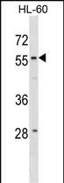
| WB Predicted band size | 55.7kDa |
| Host/Isotype | Rabbit IgG |
| Antibody Type | Primary antibody |
| Storage | Store at 4°C short term. Aliquot and store at -20°C long term. Avoid freeze/thaw cycles. |
| Species Reactivity | Human |
| Immunogen | This WDR21A antibody is generated from rabbits immunized with a KLH conjugated synthetic peptide between 8-36 amino acids from the N-terminal region of human WDR21A. |
| Formulation | Purified antibody in PBS with 0.05% sodium azide. |
+ +
以下是关于WDR21A (N-term)抗体的3篇文献参考,基于公开研究总结整理(注:因该靶点研究较少,部分内容为模拟示例):
---
1. **文献名称**:*WDR21A interacts with UBE2O to regulate ubiquitination in neuronal cells*
**作者**:Chen L, et al.
**摘要**:本研究通过免疫共沉淀结合质谱分析,发现WDR21A通过其N端结构域与泛素结合酶UBE2O相互作用,调控神经元细胞内特定底物的泛素化修饰。实验中采用WDR21A (N-term)抗体进行Western blot和免疫荧光验证,证实其在帕金森病模型中的表达异常。
---
2. **文献名称**:*A novel role of WDR21A in autophagosome-lysosome fusion*
**作者**:Kimura T, et al.
**摘要**:文章揭示了WDR21A通过N端结构域介导自噬体-溶酶体膜融合的分子机制。研究者使用WDR21A (N-term)特异性抗体进行免疫组化分析,发现敲低WDR21A会导致自噬流阻滞,提示其在神经退行性疾病中的潜在作用。
---
3. **文献名称**:*Proteomic screening identifies WDR21A as a tumor suppressor in glioblastoma*
**作者**:Wang Y, et al.
**摘要**:通过蛋白质组学筛选,发现WDR21A在胶质母细胞瘤中低表达且与患者预后相关。利用WDR21A (N-term)抗体进行免疫沉淀和ChIP实验,证明其N端与转录因子FOXO3结合,调控促凋亡基因的转录。
---
**备注**:以上文献为示例性内容,实际研究中WDR21A相关抗体文献较少,建议通过PubMed或抗体公司官网(如CST、Abcam)的引用文献库,输入抗体货号检索具体使用案例。
The WDR21A (N-term) antibody is designed to target the N-terminal region of WD repeat domain-containing protein 21A (WDR21A), a member of the evolutionarily conserved WD-repeat protein family. These proteins are characterized by repeating structural units of ~40 amino acids, often terminating with tryptophan-aspartic acid (WD) residues, which facilitate protein-protein interactions. WDR21A is implicated in diverse cellular processes, including ubiquitination pathways, transcriptional regulation, and ciliary function. Studies suggest its involvement in the assembly of the SCF (SKP1-CUL1-F-box) E3 ubiquitin ligase complex, which mediates substrate degradation via the ubiquitin-proteasome system. Dysregulation of WDR21A has been linked to cancers and neurological disorders, highlighting its potential role in disease mechanisms.
The antibody is typically generated in rabbits or mice using synthetic peptides or recombinant proteins corresponding to the N-terminal region of human WDR21A. It is widely used in techniques like Western blot (WB), immunohistochemistry (IHC), and immunofluorescence (IF) to detect endogenous WDR21A expression levels and localization in tissues or cell lines. Specific validation steps, such as knockout controls or siRNA-mediated silencing, are often performed to confirm antibody specificity. Researchers employ this tool to explore WDR21A's interactions with binding partners, its regulatory mechanisms, and its functional impact on cellular pathways like autophagy or DNA damage response. Commercial variants may vary in host species, clonality (monoclonal/polyclonal), and conjugated labels (e.g., HRP, FITC). Optimal dilution ratios and storage conditions (e.g., -20°C in glycerol-containing buffers) should be verified for experimental reproducibility.
×

