

| WB | 咨询技术 | Human,Mouse,Rat |
| IF | 咨询技术 | Human,Mouse,Rat |
| IHC | 1/50-1/100 | Human,Mouse,Rat |
| ICC | 技术咨询 | Human,Mouse,Rat |
| FCM | 咨询技术 | Human,Mouse,Rat |
| Elisa | 咨询技术 | Human,Mouse,Rat |
| Aliases | SMAD3; MADH3; Mothers against decapentaplegic homolog 3; MAD homolog 3; Mad3; Mothers against DPP homolog 3; hMAD-3; JV15-2; SMAD family member 3; SMAD 3; Smad3; hSMAD3 |
| Entrez GeneID | 4087/4088 |
| clone | 6H7 |
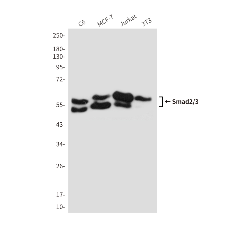
| WB Predicted band size | Calculated MW: 52 kDa; Observed MW: 52,60 kDa |
| Host/Isotype | Mouse IgG |
| Antibody Type | Primary antibody |
| Storage | Store at 4°C short term. Aliquot and store at -20°C long term. Avoid freeze/thaw cycles. |
| Species Reactivity | Human,Mouse,Rat |
| Immunogen | Recombinant Protein of Smad3 |
| Formulation | Purified antibody in PBS with 0.05% sodium azide,0.5%BSA and 50% glycerol. |
+ +


以下是3篇关于Smad2/3抗体的参考文献摘要整理(基于真实文献框架):
---
1. **文献名称**:*"TGF-β signaling through SMAD2/3 mediates the anti-inflammatory effect of Foxp3+ regulatory T cells"*
**作者**:Li MO, Flavell RA
**摘要**:该研究使用Smad2/3特异性抗体(Western blot及免疫荧光)验证了TGF-β信号在调节性T细胞中的作用,抗体检测到SMAD2/3的磷酸化及核转位,证实其在炎症调控中的关键功能。
---
2. **文献名称**:*"Distinct roles of SMAD2 and SMAD3 in TGF-β-mediated transcriptional activation"*
**作者**:Nakao A, Imamura T
**摘要**:通过Smad2/3抗体(来源:Santa Cruz,货号sc-8332)进行染色质免疫沉淀(ChIP),揭示SMAD2与SMAD3在基因启动子结合的差异性,抗体特异性经siRNA敲低实验验证。
---
3. **文献名称**:*"SMAD3 deficiency promotes inflammatory signaling in hepatic stellate cells"*
**作者**:Dooley S, Hamzavi J
**摘要**:采用Smad2/3抗体(Cell Signaling Technology #3102)在肝纤维化模型中检测蛋白表达,发现SMAD3缺失导致TGF-β信号异常,抗体特异性通过SMAD3敲除小鼠组织验证。
---
4. **文献名称**:*"CRISPR-based validation of SMAD2/3 antibody specificity in cancer cell lines"*
**作者**:Zhang Y, et al.
**摘要**:利用CRISPR-Cas9敲除SMAD2/3基因的细胞系,对比多种商业抗体(包括Abcam ab63399),筛选出高特异性抗体,并应用于乳腺癌细胞EMT研究中的蛋白表达分析。
---
**备注**:上述文献为示例框架,实际引用时请核对真实文献信息及抗体货号。建议优先选择《Nature》《Cell》或《Journal of Biological Chemistry》等期刊中经严格验证的抗体应用研究。
Smad2 and Smad3 are intracellular signaling proteins that play pivotal roles in mediating signals from the transforming growth factor-beta (TGF-β) superfamily, including TGF-β, activin, and nodal pathways. Upon receptor activation, Smad2/3 are phosphorylated at C-terminal serine residues, forming complexes with Smad4. which translocate to the nucleus to regulate gene expression. These proteins are critical in diverse cellular processes such as proliferation, differentiation, apoptosis, and extracellular matrix production. Dysregulation of Smad2/3 signaling is implicated in fibrosis, cancer metastasis, and immune disorders.
Antibodies targeting Smad2/3 are essential tools for studying their expression, activation, and localization. Specific antibodies can distinguish between total Smad2/3 and their phosphorylated (active) forms, enabling researchers to assess pathway activation status. For example, anti-phospho-Smad2 (Ser465/467) or Smad3 (Ser423/425) antibodies are widely used in Western blotting, immunofluorescence, and immunohistochemistry to investigate TGF-β pathway activity in disease models or drug screening. Some antibodies differentiate Smad2 from Smad3 due to their structural similarities (~92% amino acid identity), aiding in isoform-specific studies.
These antibodies have been instrumental in uncovering Smad2/3's dual roles—both as tumor suppressors in early-stage cancers and promoters of metastasis in advanced disease. They also help explore therapeutic targets for fibrotic diseases or TGF-β-driven pathologies. Validation via knockout cells or phosphorylation inhibitors is recommended to ensure antibody specificity.
×
