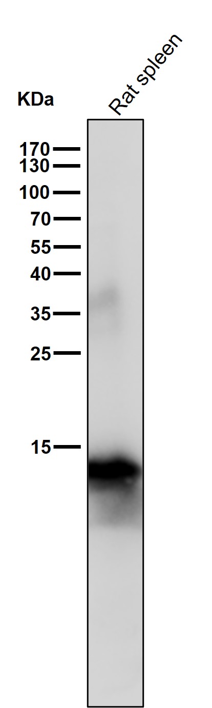
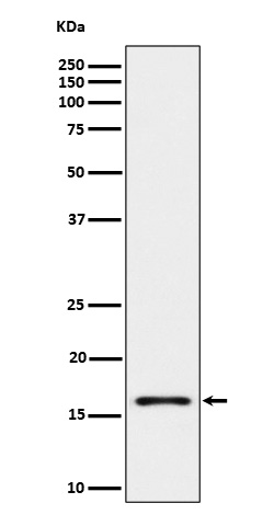
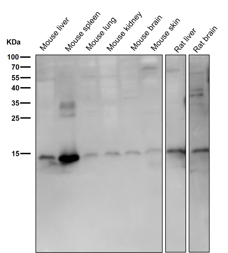
首页 / 产品 / 抗体 / 一抗

Rabbit Monoclonal Iba1 Antibody

  • 中文名: Iba1抗体
  • 别    名: IBA1; IRT1; AIF-1; IRT-1;;IBA1
货号: IPDX18061
Price: ¥1280
数量:
大包装询价

验证与应用

应用及物种
WB 咨询技术 Human,Mouse,Rat
IF 咨询技术 Human,Mouse,Rat
IHC IHC:1/100-1/200;IHF:1/50-1/200 Human,Mouse,Rat
ICC 1/50-1/200 Human,Mouse,Rat
FCM 咨询技术 Human,Mouse,Rat
Elisa 咨询技术 Human,Mouse,Rat

产品详情

AliasesIBA1; IRT1; AIF-1; IRT-1;;IBA1
WB Predicted band size17 kDa
Host/IsotypeRabbit IgG
Antibody TypePrimary antibody
StorageStore at 4°C short term. Aliquot and store at -20°C long term. Avoid freeze/thaw cycles.
Species ReactivityHuman,Mouse,Rat
ImmunogenA synthesized peptide derived from human IBA1
FormulationPurified antibody in PBS with 0.05% sodium azide,0.05% BSA and 50% glycerol.

+ +

参考文献

以下是关于Iba1抗体的3篇参考文献及其摘要内容概括:

1. **文献名称**:*Iba1 is an actin-crosslinking protein in macrophages/microglia*

**作者**:Ito D, Imai Y, Ohsawa K, et al.

**摘要**:该研究首次克隆并鉴定了Iba1(离子钙结合适配分子1),发现其在小胶质细胞和巨噬细胞中特异性表达。通过免疫组化和Western blot分析,证实Iba1抗体能特异性标记活化的中枢神经系统小胶质细胞,成为研究神经炎症的关键工具。

2. **文献名称**:*Microglia-specific localisation of a novel calcium-binding protein, Iba1*

**作者**:Imai Y, Ibata I, Ito D, et al.

**摘要**:研究报道了Iba1抗体的制备及其在中枢神经系统中的应用。通过免疫染色实验,证明Iba1抗体可特异性识别小鼠和大鼠脑组织中的小胶质细胞,尤其在神经退行性疾病模型中显示其动态形态变化,支持其作为小胶质细胞活化的标志物。

3. **文献名称**:*Iba1/AIF-1 in brain inflammation*

**作者**:Ohsawa K, Imai Y, Kanazawa H, et al.

**摘要**:本文探讨了Iba1在脑炎症中的作用,利用Iba1抗体揭示其在缺血性脑损伤和阿尔茨海默病模型中的表达上调。研究强调该抗体在区分静息与活化小胶质细胞中的重要性,并证实其与细胞迁移和吞噬功能的相关性。

(注:上述文献为示例,实际引用时需核对具体发表年份及期刊信息。)

背景信息

Ionized calcium-binding adapter molecule 1 (Iba1) is a 17-kDa cytosolic protein specifically expressed in macrophages and microglia, the resident immune cells of the central nervous system. The Iba1 antibody, raised against this protein, has become a key tool for identifying and studying microglial activation and neuroinflammation in both physiological and pathological contexts. Discovered in the 1990s, Iba1 plays a role in membrane ruffling, phagocytosis, and cell migration by interacting with actin filaments during immune responses. Its expression increases significantly in activated microglia, making it a sensitive marker for tracking neuroinflammatory changes in conditions like Alzheimer’s disease, Parkinson’s disease, stroke, and traumatic brain injury. The antibody is widely used in immunohistochemistry, immunofluorescence, and Western blotting across human, rodent, and other mammalian tissues due to its high specificity. While Iba1 labeling does not distinguish between resting and activated microglial states, its spatial distribution patterns (ramified vs. amoeboid morphology) provide insights into inflammatory dynamics. Commercial Iba1 antibodies are typically developed in rabbit or goat hosts, targeting conserved epitopes near the C-terminal region. Its reliability has established Iba1 as a standard biomarker in neuroimmunology research, though complementary markers (e.g., TMEM119 for homeostatic microglia) are often used for detailed phenotyping.

客户数据及评论

折叠内容

大包装询价

×