
| WB | 咨询技术 | Human,Mouse,Rat |
| IF | ChIP:1/20-1/50 | Human,Mouse,Rat |
| IHC | IHC:1/100-1/200;IHF:1/50-1/200 | Human,Mouse,Rat |
| ICC | 1/50-1/200 | Human,Mouse,Rat |
| FCM | 咨询技术 | Human,Mouse,Rat |
| Elisa | 咨询技术 | Human,Mouse,Rat |
| Aliases | Histone H3.1, Histone H3, HIST1H3A;;p-Histone H3 (S11) |
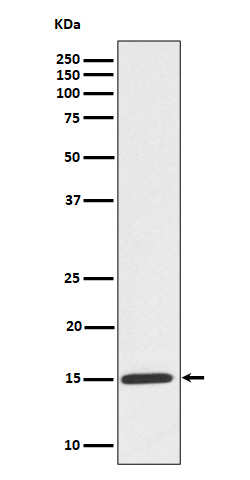
| WB Predicted band size | 15 kDa |
| Host/Isotype | Rabbit IgG |
| Antibody Type | Primary antibody |
| Storage | Store at 4°C short term. Aliquot and store at -20°C long term. Avoid freeze/thaw cycles. |
| Species Reactivity | Human,Mouse,Rat |
| Immunogen | A synthesized peptide derived from human Histone H3.1 around the phosphorylation site of S11 |
| Formulation | Purified antibody in PBS with 0.05% sodium azide,0.05% BSA and 50% glycerol. |
+ +
以下是关于Phospho-Histone H3 (Ser10)抗体的3篇参考文献,涵盖其在细胞周期研究、肿瘤检测及神经科学中的应用:
---
1. **文献名称**:*Phosphorylation of histone H3 is required for chromosome condensation and mitotic progression*
**作者**:Hendzel, M.J. et al.
**摘要**:该研究首次揭示了组蛋白H3第10位丝氨酸(S10)的磷酸化与有丝分裂期间染色体凝缩的直接关联。通过免疫荧光和抗体特异性检测,证实H3S10磷酸化是细胞进入有丝分裂的关键标志,并参与调控染色体结构的动态变化。
---
2. **文献名称**:*Histone H3 phosphorylation and expression of cyclins A and B1 measured in individual cells during their progression through G2 and mitosis*
**作者**:Juan, G. et al.
**摘要**:作者利用Phospho-Histone H3 (S10)抗体结合流式细胞术,定量分析了单个细胞在G2/M期的周期蛋白(Cyclins)表达与H3磷酸化的关系。该研究建立了H3S10磷酸化作为有丝分裂特异性标记物的技术方法,并用于评估肿瘤细胞增殖活性。
---
3. **文献名称**:*Mitosis-specific phosphorylation of histone H3 initiates primarily within pericentromeric heterochromatin during G2 and spreads in an ordered fashion coincident with mitotic chromosome condensation*
**作者**:Wei, Y. et al.
**摘要**:研究通过Phospho-H3 (S10)抗体的免疫定位,发现H3磷酸化起始于G2期的着丝粒周围异染色质区域,并随染色体凝缩逐步扩散。该发现揭示了磷酸化时空动态与有丝分裂进程的分子机制关联,为癌症病理中异常细胞增殖的诊断提供了依据。
---
如需更多文献或特定领域应用,可进一步补充。
The Phospho-Histone H3 (Ser10) antibody detects histone H3 phosphorylated at serine 10. a post-translational modification tightly linked to chromatin condensation during mitosis. Histone H3. a core component of nucleosomes, undergoes dynamic phosphorylation during the cell cycle, particularly at the G2/M transition. This modification, mediated by Aurora kinases and other mitotic regulators, is a hallmark of mitotic chromatin and plays a role in chromosome segregation.
The antibody is widely used as a marker for mitotic cells in research, as phosphorylation at Ser10 peaks during mitosis and rapidly declines upon exit. It enables precise identification of cells in the M phase, making it valuable in studies of cell proliferation, cancer biology (e.g., assessing tumor aggressiveness), neurogenesis, and developmental processes. Common applications include immunofluorescence, immunohistochemistry, and Western blotting to visualize mitotic cells in tissues or cultured samples. It is also employed in flow cytometry to quantify mitotic indices in drug response studies or cell cycle analyses.
Due to its specificity for the phosphorylated epitope, this antibody helps distinguish mitotic cells from those in other cell cycle phases or apoptotic states. Cross-reactivity has been validated in multiple species, including humans, mice, and rats. Its utility extends to pathological evaluations, such as grading proliferative activity in tumors, and mechanistic studies exploring cell cycle deregulation in diseases or genetic models.
×

