
| WB | 咨询技术 | Human,Mouse,Rat |
| IF | 1/20-1/50 | Human,Mouse,Rat |
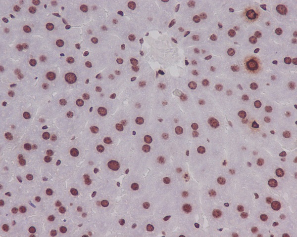
| IHC | IHC:1/100-1/200;IHF:1/50-1/200 | Human,Mouse,Rat |
| ICC | 1/50-1/200 | Human,Mouse,Rat |
| FCM | 咨询技术 | Human,Mouse,Rat |
| Elisa | 咨询技术 | Human,Mouse,Rat |
| Aliases | H3 histone, family 3A; H3 histone, family 3B (H3.3B); H3.3A; H3.3B; H33; H3F3; H3F3A; H3F3B; Histone H3.3;;p-Histone H3 (S11) |
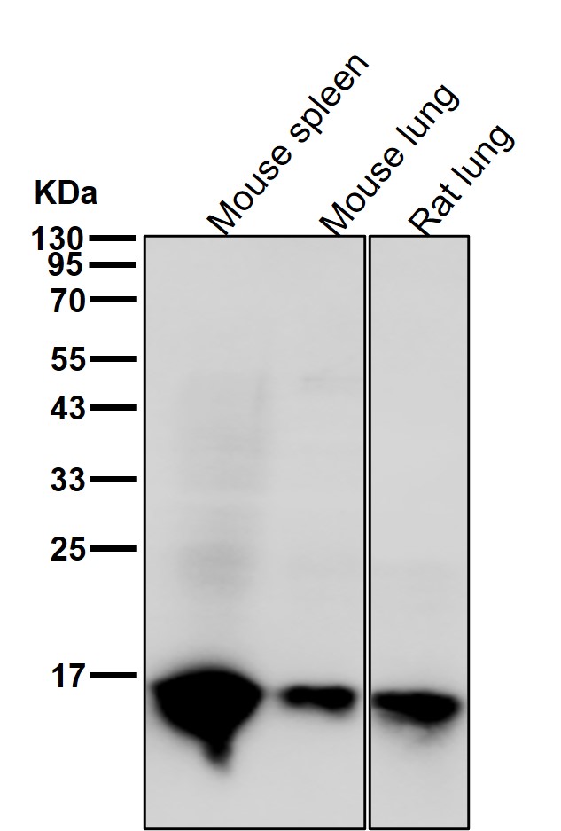
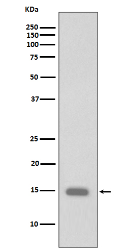
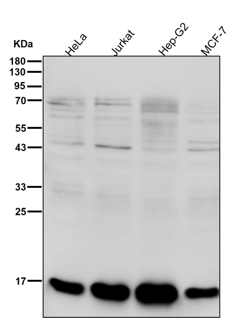
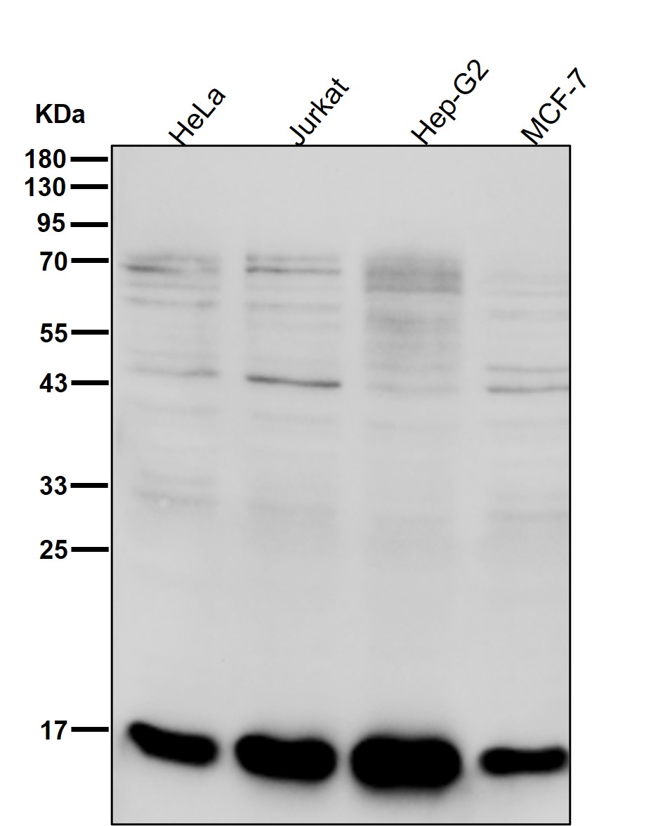
| WB Predicted band size | 15 kDa |
| Host/Isotype | Rabbit IgG |
| Antibody Type | Primary antibody |
| Storage | Store at 4°C short term. Aliquot and store at -20°C long term. Avoid freeze/thaw cycles. |
| Species Reactivity | Human,Mouse,Rat |
| Immunogen | A synthesized peptide derived from human Histone H3.1 around the phosphorylation site of S11 |
| Formulation | Purified antibody in PBS with 0.05% sodium azide,0.05% BSA and 50% glycerol. |
+ +


以下是关于Phospho-Histone H3 (S10)抗体的3篇参考文献及其摘要概括:
---
1. **文献名称**:*Phosphorylation of histone H3 at serine 10 correlates with chromosome condensation during mitosis and meiosis*
**作者**:Hendzel, M.J. 等
**摘要**:该研究通过免疫荧光和Western blot分析,揭示了组蛋白H3在Ser10位点的磷酸化与哺乳动物细胞有丝分裂及减数分裂中染色体凝聚的直接关联,提出其作为细胞周期G2/M期转变的关键表观遗传标记。
2. **文献名称**:*Mitotic-specific phosphorylation of histone H3 initiates primarily from pericentromeric heterochromatin*
**作者**:Crosio, C. 等
**摘要**:研究利用Phospho-Histone H3 (S10)抗体发现,哺乳动物细胞中H3的Ser10磷酸化起始于着丝粒周围异染色质区域,并依赖Aurora激酶活性,提示其在染色体分离和细胞周期调控中的关键作用。
3. **文献名称**:*Histone H3 phosphorylation and expression of cyclins A and B1 measured in individual cells during their progression through G2 and mitosis*
**作者**:Juan, G. 等
**摘要**:通过流式细胞术和多色荧光成像,该文献证明H3(S10)磷酸化水平与细胞周期蛋白Cyclin B1表达同步升高,可作为有丝分裂进程的高灵敏度标记,用于肿瘤细胞增殖活性评估。
---
这些文献涵盖了该抗体在染色体动态、激酶调控及肿瘤研究中的应用,均为领域内经典研究。如需扩展,可进一步检索近年研究其在特定疾病模型(如癌症或干细胞分化)中的新进展。
The Phospho-Histone H3 (Ser10) antibody is a critical tool for studying mitotic progression and chromatin dynamics. Histone H3 is a core component of nucleosomes, and its phosphorylation at serine 10 (S10) is tightly associated with chromosome condensation during mitosis. This post-translational modification peaks during the G2/M phase of the cell cycle, serving as a hallmark of mitotic cells. The antibody specifically detects this phosphorylation event, enabling researchers to visualize and quantify cells actively undergoing division.
Widely used in immunofluorescence, flow cytometry, and Western blotting, this antibody helps identify mitotic cells in tissues, cultured cells, or experimental models. Its applications extend to cancer research, where it aids in assessing tumor proliferation rates, as highly aggressive cancers often exhibit elevated phospho-Histone H3 (S10) levels. It also plays a role in developmental biology and drug discovery, particularly in screening compounds that modulate cell cycle progression.
The antibody's specificity is validated using knockout controls or phosphatase-treated samples to confirm signal loss. Proper sample preparation, including formaldehyde fixation and avoidance of harsh detergents, is crucial to preserve the phosphorylation epitope. By providing a direct readout of mitotic activity, the Phospho-Histone H3 (S10) antibody remains indispensable for understanding cell division mechanisms, disease pathology, and therapeutic interventions targeting proliferation.
×
