


| WB | 咨询技术 | Human,Mouse,Rat |
| IF | 咨询技术 | Human,Mouse,Rat |
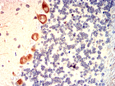
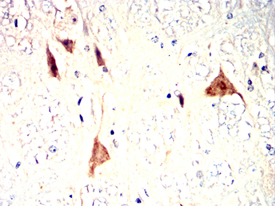
| IHC | 1/50-1/100 | Human,Mouse,Rat |
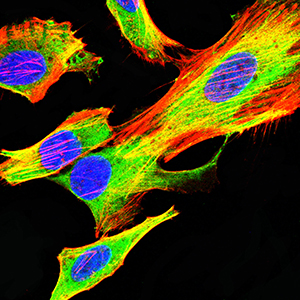
| ICC | 1/50-1/100 | Human,Mouse,Rat |
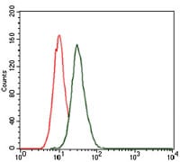
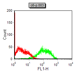
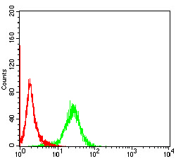
| FCM | 1/200 - 1/400 | Human,Mouse,Rat |
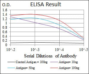
| Elisa | 1/10000 | Human,Mouse,Rat |
| Aliases | LC3B; ATG8F; MAP1A/1BLC3 |
| Entrez GeneID | 81631 |
| clone | 5H12 |
| WB Predicted band size | 14.6kDa |
| Host/Isotype | Mouse IgG1 |
| Antibody Type | Primary antibody |
| Storage | Store at 4°C short term. Aliquot and store at -20°C long term. Avoid freeze/thaw cycles. |
| Species Reactivity | Human,Mouse,Rat,Monkey |
| Immunogen | Purified recombinant fragment of human MAP1LC3B expressed in E. Coli. |
| Formulation | Ascitic fluid containing 0.03% sodium azide. |
+ +


以下是关于MAP1LC3B抗体的3篇代表性文献的简要总结:
1. **文献名称**:*LC3. a mammalian homologue of yeast Apg8p, is localized in autophagosome membranes after processing*
**作者**:Kabeya, Y. et al.
**摘要**:该研究首次鉴定并描述了哺乳动物LC3蛋白(包括LC3B)的功能,阐明了其在自噬体形成中的关键作用。作者开发了特异性识别LC3B的抗体,验证了其在免疫印迹(Western blot)和免疫荧光中的应用,证明了LC3B-II(脂化形式)作为自噬体标志物的可靠性。
2. **文献名称**:*Guidelines for the use and interpretation of assays for monitoring autophagy*
**作者**:Klionsky, D.J. et al.
**摘要**:这篇权威指南汇总了自噬研究的标准化方法,其中专门讨论了LC3B抗体的应用场景(如Western blot检测LC3B-I/II转化、免疫荧光定位自噬体)。文献强调了不同抗体(如抗N端或C端抗体)的适用性差异,并提醒注意假阳性结果的潜在问题。
3. **文献名称**:*Dissection of the autophagosome maturation process by a novel reporter protein, tandem fluorescent-tagged LC3*
**作者**:Mizushima, N. et al.
**摘要**:研究通过构建串联荧光标记的LC3B(mRFP-GFP-LC3)探针,开发了动态监测自噬流的方法。文中对比了多种商业LC3B抗体的特异性,验证了其在流式细胞术和共聚焦显微镜中的有效性,为抗体选择提供了实验依据。
4. **文献名称**:*LC3 and Autophagy*
**作者**:Tanida, I. et al.
**摘要**:该综述系统总结了LC3蛋白家族(包括LC3B)在自噬中的分子机制,并详细分析了不同LC3B抗体的表位特异性及其适用实验类型(如免疫组化需选择识别天然构象的抗体)。
**注**:以上文献均发表于*Autophagy*等高水平期刊,可作为LC3B抗体使用及自噬研究的核心参考文献。
The MAP1LC3B (microtubule-associated protein 1 light chain 3 beta) antibody is a critical tool for studying autophagy, a cellular degradation process essential for maintaining homeostasis. LC3B, a mammalian homolog of yeast Atg8. is a key marker of autophagosomes. During autophagy, cytosolic LC3B (LC3B-I) conjugates to phosphatidylethanolamine, forming LC3B-II, which integrates into autophagosomal membranes. This conversion is widely used to monitor autophagic activity.
The antibody specifically detects LC3B isoforms, distinguishing between LC3B-I (18 kDa) and LC3B-II (16 kDa) via Western blotting. It is also employed in immunofluorescence (IF) and immunohistochemistry (IHC) to visualize autophagosome formation in cells or tissues. Researchers utilize it to explore autophagy's role in diseases like cancer, neurodegenerative disorders, and infections, as dysregulated autophagy contributes to these conditions.
Commercial LC3B antibodies are typically raised against synthetic peptides corresponding to conserved regions of human LC3B, ensuring cross-reactivity with homologs in mice, rats, and other species. However, interpretation requires caution: LC3B-II levels alone may not fully represent autophagic flux, necessitating complementary assays (e.g., lysosomal inhibition). Variability in antibody specificity and batch consistency also underscores the need for validation using LC3B-knockout controls. Overall, the LC3B antibody remains indispensable for elucidating autophagy mechanisms in health and disease.
×
