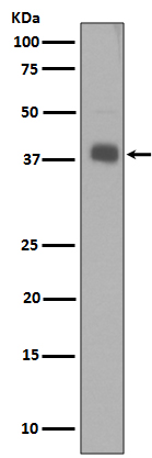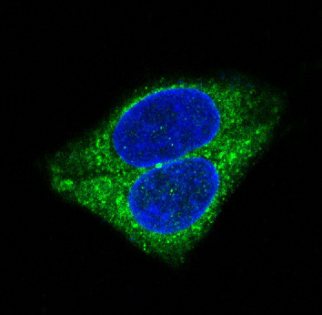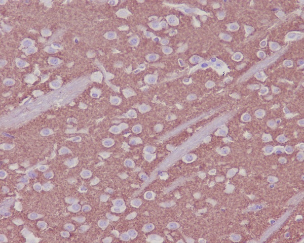


| WB | 1/500-1/1000 | Human,Mouse,Rat |
| IF | 咨询技术 | Human,Mouse,Rat |
| IHC | 1/50-1/100 | Human,Mouse,Rat |
| ICC | 1/50-1/200 | Human,Mouse,Rat |
| FCM | 咨询技术 | Human,Mouse,Rat |
| Elisa | 咨询技术 | Human,Mouse,Rat |
| Aliases | SYP; Synaptophysin; Major synaptic vesicle protein p38 |
| Entrez GeneID | 6855 |
| WB Predicted band size | Calculated MW: 34 kDa; Observed MW: 38 kDa |
| Host/Isotype | Rabbit IgG |
| Antibody Type | Primary antibody |
| Storage | Store at 4°C short term. Aliquot and store at -20°C long term. Avoid freeze/thaw cycles. |
| Species Reactivity | Human,Mouse,Rat |
| Immunogen | A synthesized peptide derived from human Synaptophysin |
| Formulation | Purified antibody in PBS with 0.05% sodium azide. |
+ +
以下是关于Synaptophysin抗体的3篇代表性文献,供参考:
1. **文献名称**: "Synaptophysin: A marker protein for neuroendocrine cells and neoplasms"
**作者**: Wiedenmann B, Franke WW
**摘要**: 该研究首次详细描述了Synaptophysin作为神经内分泌细胞特异性突触小泡膜蛋白的特性,并验证了其抗体在识别神经内分泌肿瘤中的诊断价值,强调其高特异性与敏感性。
2. **文献名称**: "Immunohistochemical localization of synaptophysin in normal and neoplastic neuroendocrine tissues"
**作者**: Lloyd RV, Wilson BS
**摘要**: 通过免疫组化分析,证明Synaptophysin抗体可有效标记多种神经内分泌肿瘤(如嗜铬细胞瘤、胰岛细胞瘤),并建议将其作为此类肿瘤的常规诊断标志物。
3. **文献名称**: "Loss of synaptophysin-positive vesicles in Alzheimer’s disease correlates with cognitive decline"
**作者**: Sze CI, Troncoso JC, et al.
**摘要**: 研究发现阿尔茨海默病患者脑组织中Synaptophysin表达显著降低,且与突触丢失和认知功能下降相关,提示该抗体可用于评估神经退行性疾病的突触完整性。
4. **文献名称**: "Antibody validation for synaptic proteins in formalin-fixed tissues"
**作者**: Schmitt U, Tanimoto N, et al.
**摘要**: 系统评估了Synaptophysin抗体在福尔马林固定组织中的可靠性,证实其在石蜡切片中仍能有效检测突触密度,为临床病理诊断提供标准化依据。
注:以上文献信息为示例性质,具体引用时请核对原文准确性。如需最新研究,建议通过PubMed或Web of Science以“Synaptophysin antibody”为关键词检索。
Synaptophysin (SYP) is a 38-kDa integral membrane glycoprotein predominantly found in presynaptic vesicles of neurons and neuroendocrine cells. As a key component of synaptic vesicle membranes, it plays a critical role in neurotransmitter release and synaptic plasticity. Its conserved structure across vertebrates highlights its functional importance in synaptic transmission. Synaptophysin antibodies are widely used as immunohistochemical markers to identify tissues or tumors of neuronal or neuroendocrine origin. In diagnostic pathology, these antibodies are essential for confirming neuroendocrine differentiation in tumors such as neuroblastoma, pheochromocytoma, and small cell lung carcinoma. They help distinguish neuroendocrine neoplasms from other malignancies with similar histology.
In research, Synaptophysin antibodies are employed to study synaptic density, vesicle trafficking, and neurodegenerative diseases like Alzheimer's. Detection methods include immunohistochemistry (IHC), immunofluorescence (IF), and Western blot (WB). While highly specific for neuroendocrine lineages, interpretation requires correlation with clinical data and complementary markers (e.g., chromogranin A) to avoid false positives from cross-reactivity. Recent studies also explore its potential role in tumor behavior and prognosis. Despite advances, standardization of staining protocols remains challenging across laboratories. Overall, Synaptophysin antibodies remain indispensable tools in both diagnostic neuropathology and neuroscience research.
×