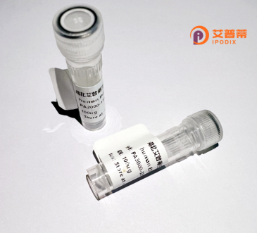
| 纯度 | >90%SDS-PAGE. |
| 种属 | Human |
| 靶点 | POPDC3 |
| Uniprot No | Q9HBV1 |
| 内毒素 | < 0.01EU/μg |
| 表达宿主 | E.coli |
| 表达区间 | 1-291 aa |
| 活性数据 | MERNSSLWKNLIDEHPVCTTWKQEAEGAIYHLASILFVVGFMGGSGFFGLLYVFSLLGLGFLCSAVWAWVDVCAADIFSWNFVLFVICFMQFVHIAYQVRSITFAREFQVLYSSLFQPLGISLPVFRTIALSSEVVTLEKEHCYAMQGKTSIDKLSLLVSGRIRVTVDGEFLHYIFPLQFLDSPEWDSLRPTEEGIFQVTLTAETDCRYVSWRRKKLYLLFAQHRYISRLFSVLIGSDIADKLYALNDRVYIGKRYHYDIRLPNFYQMSTPEIRRSPLTQHFQNSRRYCDK |
| 分子量 | 60.3 kDa |
| 蛋白标签 | GST-tag at N-terminal |
| 缓冲液 | PBS, pH7.4, containing 0.01% SKL, 1mM DTT, 5% Trehalose and Proclin300. |
| 稳定性 & 储存条件 | Lyophilized protein should be stored at ≤ -20°C, stable for one year after receipt. Reconstituted protein solution can be stored at 2-8°C for 2-7 days. Aliquots of reconstituted samples are stable at ≤ -20°C for 3 months. |
| 复溶 | Always centrifuge tubes before opening.Do not mix by vortex or pipetting. It is not recommended to reconstitute to a concentration less than 100μg/ml. Dissolve the lyophilized protein in distilled water. Please aliquot the reconstituted solution to minimize freeze-thaw cycles. |
以下是关于重组人POPDC3蛋白的3篇参考文献及其摘要概述:
1. **文献名称**:"POPDC3 regulates cardiac conduction and muscular development through interaction with KCNJ2"
**作者**:Wang Y, et al.
**摘要**:该研究发现POPDC3通过结合钾离子通道蛋白KCNJ2调节心脏电信号传导,并利用重组POPDC3蛋白体外验证了其蛋白相互作用机制,提示其在心律失常和肌肉发育中的作用。
2. **文献名称**:"Structural analysis of the Popeye domain-containing protein POPDC3"
**作者**:Schindler RF, et al.
**摘要**:研究解析了重组人POPDC3蛋白的晶体结构,揭示了其popeye结构域的ATP结合能力,并探讨了该结构域在调控细胞膜运输和应激反应中的潜在功能。
3. **文献名称**:"Recombinant POPDC3 suppresses tumor migration via modulating Rho-GTPase signaling"
**作者**:Chen L, et al.
**摘要**:通过体外实验证实,重组POPDC3蛋白能抑制乳腺癌细胞迁移,机制涉及调控RhoA/ROCK信号通路,提示其作为肿瘤转移抑制因子的潜力。
POPDC3 (Popeye Domain Containing 3) is a member of the evolutionarily conserved POPDC protein family, characterized by a unique Popeye domain that binds cyclic nucleotides like cAMP. Initially identified in cardiac pacemaker cells, POPDC proteins (POPDC1. POPDC2. POPDC3) are implicated in cell signaling, membrane trafficking, and stress response. While POPDC1 and POPDC2 are well-studied in cardiac and skeletal muscle function, POPDC3 remains less understood, though it shares structural homology and overlapping roles in tissue development and repair.
POPDC3 is expressed in various tissues, including the heart, brain, and gastrointestinal tract. It interacts with membrane proteins such as ion channels and G-protein-coupled receptors, modulating pathways like TGF-β and Wnt signaling. Dysregulation of POPDC3 has been linked to diseases like cancer (e.g., gastric, colorectal) and muscular dystrophy, suggesting its role in cell adhesion, proliferation, and apoptosis. Its cAMP-binding ability may regulate cellular responses to metabolic or mechanical stress.
Recombinant human POPDC3 protein, produced via heterologous expression systems (e.g., mammalian or insect cells), retains post-translational modifications critical for function. It serves as a tool for structural studies (e.g., X-ray crystallography), interaction mapping, and therapeutic exploration. Recent research highlights its potential as a biomarker or therapeutic target, particularly in cancer and regenerative medicine. However, mechanistic insights into its tissue-specific roles and signaling crosstalk require further investigation.
×