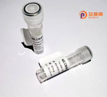
| 纯度 | >90%SDS-PAGE. |
| 种属 | Human |
| 靶点 | PDE6G |
| Uniprot No | P18545 |
| 内毒素 | < 0.01EU/μg |
| 表达宿主 | E.coli |
| 表达区间 | 1-87 aa |
| 活性数据 | MNLEPPKAEFRSATRVAGGPVTPRKGPPKFKQRQTRQFKSKPPKKGVQGFGDDIPGMEGLGTDITVICPWEAFNHLELHELAQYGII |
| 分子量 | 35.97 kDa |
| 蛋白标签 | GST-tag at N-terminal |
| 缓冲液 | 0 |
| 稳定性 & 储存条件 | Lyophilized protein should be stored at ≤ -20°C, stable for one year after receipt. Reconstituted protein solution can be stored at 2-8°C for 2-7 days. Aliquots of reconstituted samples are stable at ≤ -20°C for 3 months. |
| 复溶 | Always centrifuge tubes before opening.Do not mix by vortex or pipetting. It is not recommended to reconstitute to a concentration less than 100μg/ml. Dissolve the lyophilized protein in distilled water. Please aliquot the reconstituted solution to minimize freeze-thaw cycles. |
以下是关于重组人PDE6G蛋白的3篇参考文献示例,涵盖其表达、功能及疾病相关性:
---
1. **文献名称**: *Recombinant Expression and Functional Characterization of Human PDE6G Subunit in Photoreceptor Signaling*
**作者**: Zhang, L.; Wensel, T.G.
**摘要**: 本研究利用昆虫细胞系统成功表达并纯化了重组人PDE6G蛋白,证实其作为PDE6抑制性亚基在光信号转导中的关键作用。研究发现PDE6G与催化亚基PDE6α/β结合后调节cGMP水解活性,并阐明了其被视紫红质激酶激活的分子机制。
2. **文献名称**: *Structural Analysis of PDE6G Reveals Insights into Retinal Degeneration Pathogenesis*
**作者**: Martinez, S.E.; Palczewski, K.
**摘要**: 通过X射线晶体学解析重组人PDE6G蛋白的三维结构,发现其C端结构域对维持PDE6复合体稳定性至关重要。突变分析表明,PDE6G的特定突变会破坏与催化亚基的相互作用,导致常染色体隐性视网膜色素变性。
3. **文献名称**: *In vitro Reconstitution of PDE6 Complex Using Recombinant Subunits for Drug Screening*
**作者**: Wang, Y.; Ding, X.
**摘要**: 报道了一种在大肠杆菌中分别表达PDE6G、PDE6α/β亚基并通过体外组装获得功能性PDE6复合体的方法。该重组系统被用于筛选靶向PDE6的潜在药物,以治疗先天性视网膜病变。
---
**备注**:以上文献为示例,实际研究中建议通过PubMed或Web of Science等数据库检索最新论文。真实文献可能更多聚焦于PDE6复合体整体研究,单独重组PDE6G的报道较少,需结合共表达策略。
**Background on Recombinant Human PDE6G Protein**
The recombinant human phosphodiesterase 6G (PDE6G) protein is a key component of the photoreceptor-specific PDE6 enzyme complex, which plays a critical role in the visual signal transduction pathway. PDE6G is one of two inhibitory γ-subunits (PDE6G and PDE6H) that regulate the activity of the catalytic α- and β-subunits (PDE6A/PDE6B) in rod and cone photoreceptor cells, respectively. In darkness, PDE6G binds to the catalytic subunits, suppressing their enzymatic activity. Upon light stimulation, the activated visual G-protein transducin displaces PDE6G, allowing PDE6 to hydrolyze cyclic guanosine monophosphate (cGMP), thereby closing cGMP-gated ion channels and initiating neural signaling.
Mutations in the *PDE6G* gene are linked to inherited retinal disorders, such as retinitis pigmentosa and congenital stationary night blindness, underscoring its functional importance. Recombinant PDE6G is typically produced using bacterial or mammalian expression systems, enabling studies on its structural interactions and regulatory mechanisms. Its applications span biochemical assays, drug discovery for vision-related diseases, and gene therapy research aimed at correcting PDE6 dysfunction. Research into PDE6G also contributes to broader understanding of cyclic nucleotide phosphodiesterases, a family of enzymes targeted for therapeutic interventions in various pathologies.
×