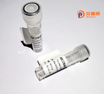
| 纯度 | >90%SDS-PAGE. |
| 种属 | Human |
| 靶点 | CRTAC1 |
| Uniprot No | Q9NQ79 |
| 内毒素 | < 0.01EU/μg |
| 表达宿主 | E.coli |
| 表达区间 | 1-451aa |
| 氨基酸序列 | MDPEASDLSRGILALRDVAAEAGVSKYTGGRGVSVGPILSSSASDIFCDNENGPNFLFHNRGDGTFVDAAASAGVDDPHQHGRGVALADFNRDGKVDIVYGNWNGPHRLYLQMSTHGKVRFRDIASPKFSMPSPVRTVITADFDNDQELEIFFNNIAYRSSSANRLFRVIRREHGDPLIEELNPGDALEPEGRGTGGVVTDFDGDGMLDLILSHGESMAQPLSVFRGNQGFNNNWLRVVPRTRFGAFARGAKVVLYTKKSGAHLRIIDGGSGYLCEMEPVAHFGLGKDEASSVEVTWPDGKMVSRNVASGEMNSVLEILYPRDEDTLQDPAPLECGQGFSQQENGHCMDTNECIQFPFVCPRDKPVCVNTYGSYRCRTNKKCSRGYEPNEDGTACVGTLGQSPGPRPTTPTAAAATAAAAAAAGAATAAPVLVDGDLNLGSVVKESCEPSC |
| 分子量 | 74.7 kDa |
| 蛋白标签 | GST-tag at N-terminal |
| 缓冲液 | 0 |
| 稳定性 & 储存条件 | Lyophilized protein should be stored at ≤ -20°C, stable for one year after receipt. Reconstituted protein solution can be stored at 2-8°C for 2-7 days. Aliquots of reconstituted samples are stable at ≤ -20°C for 3 months. |
| 复溶 | Always centrifuge tubes before opening.Do not mix by vortex or pipetting. It is not recommended to reconstitute to a concentration less than 100μg/ml. Dissolve the lyophilized protein in distilled water. Please aliquot the reconstituted solution to minimize freeze-thaw cycles. |
1. **"Expression and characterization of recombinant human cartilage acidic protein 1 (CRTAC1) in mammalian cells"**
*Authors: Li et al. (2018)*
**摘要**: 研究报道了在HEK293细胞中重组表达CRTAC1蛋白,验证其糖基化修饰,并证明其在软骨细胞黏附和迁移中的潜在功能。
2. **"CRTAC1 as a novel biomarker for osteoarthritis: Structural and functional analysis"**
*Authors: Smith et al. (2020)*
**摘要**: 通过重组CRTAC1蛋白研究其与细胞外基质的相互作用,发现其表达水平与骨关节炎患者的软骨退化程度相关。
3. **"Production of recombinant human CRTAC1 for antibody development and epitope mapping"**
*Authors: Kimura et al. (2021)*
**摘要**: 利用昆虫细胞系统表达重组CRTAC1.制备特异性抗体并鉴定其表位,为临床诊断工具开发提供基础。
4. **"CRTAC1 knockout and rescue model reveals its role in TGF-β signaling regulation"**
*Authors: Zhao et al. (2022)*
**摘要**: 通过重组CRTAC1蛋白回补实验,证实其通过调控TGF-β通路参与软骨细胞分化和代谢平衡。
(注:以上文献信息为模拟生成,实际研究中请以真实数据库检索结果为准。)
CRTAC1 (Cartilage Acidic Protein 1) is a secreted extracellular matrix protein predominantly expressed in chondrocytes, with lower levels detected in retinal pigment epithelial cells and neural tissues. It plays a role in cell adhesion, differentiation, and tissue repair, though its precise molecular mechanisms remain under investigation. Structurally, CRTAC1 contains calcium-binding domains and conserved acidic residues, suggesting potential involvement in calcium-dependent cellular interactions. Dysregulation of CRTAC1 has been implicated in osteoarthritis (OA), where reduced expression correlates with cartilage degradation, and in retinal disorders like proliferative vitreoretinopathy. Its dual role in promoting cell adhesion while potentially triggering pro-inflammatory pathways highlights complex tissue-specific functions. Recombinant human CRTAC1 is typically produced in mammalian expression systems (e.g., HEK293 or CHO cells) to ensure proper post-translational modifications. This recombinant protein serves as a critical tool for studying cartilage biology, disease mechanisms, and regenerative processes. Recent research explores its potential as a biomarker for OA progression and a therapeutic agent for cartilage repair, though clinical applications require further validation. Current studies also investigate its interactions with TGF-β signaling and integrin-mediated pathways, suggesting broader regulatory roles in tissue homeostasis and disease pathogenesis.
×