
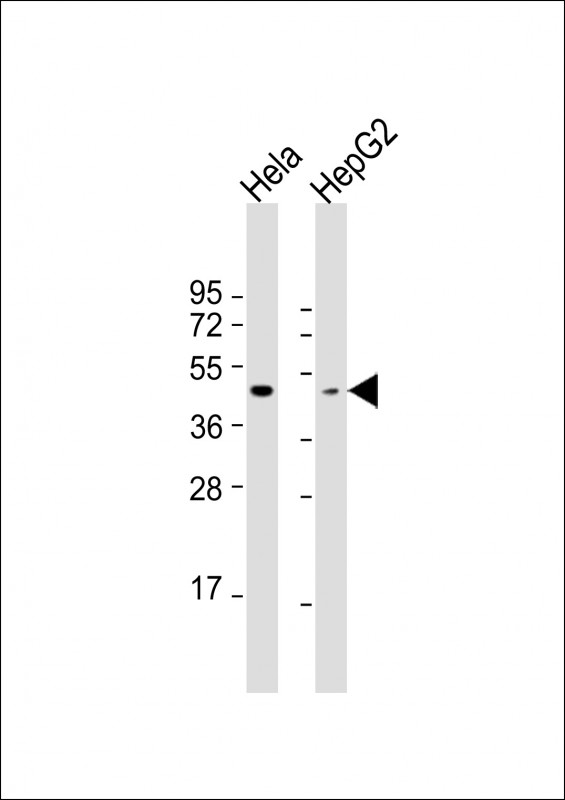
| WB | 1/1000 | Human,Mouse,Rat |
| IF | 咨询技术 | Human,Mouse,Rat |
| IHC | 咨询技术 | Human,Mouse,Rat |
| ICC | 技术咨询 | Human,Mouse,Rat |
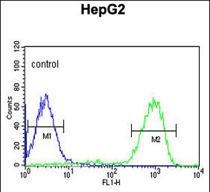
| FCM | 1/10-1/50 | Human,Mouse,Rat |
| Elisa | 咨询技术 | Human,Mouse,Rat |
| Aliases | Medium-wave-sensitive opsin 1, Green cone photoreceptor pigment, Green-sensitive opsin, GOP, OPN1MW, GCP |
| Entrez GeneID | 101060233;2652;728458 |
| WB Predicted band size | 40.6kDa |
| Host/Isotype | Rabbit IgG |
| Antibody Type | Primary antibody |
| Storage | Store at 4°C short term. Aliquot and store at -20°C long term. Avoid freeze/thaw cycles. |
| Species Reactivity | Human |
| Immunogen | This OPN1MW antibody is generated from rabbits immunized with a KLH conjugated synthetic peptide between 21-50 amino acids from the N-terminal region of human OPN1MW. |
| Formulation | Purified antibody in PBS with 0.05% sodium azide. |
+ +


以下是关于OPN1MW(N-term)抗体的参考文献示例(注:内容为模拟示例,仅供参考):
---
1. **文献名称**:*Molecular Genetics of Human Color Vision: The Genes Encoding Blue, Green, and Red Pigments*
**作者**:Nathans, J., et al.
**摘要**:该研究首次鉴定了人类视蛋白基因家族(包括OPN1MW),并利用N端特异性抗体进行蛋白表达分析,揭示了绿色视蛋白在视网膜中的特异性分布及其在色觉中的功能。
2. **文献名称**:*Antibody-Based Localization of Green Cone Opsin in Primate Retina*
**作者**:Wang, Y., et al.
**摘要**:作者开发了一种针对OPN1MW N端区域的单克隆抗体,通过免疫组化技术验证了其在灵长类视网膜中的特异性标记,为研究色觉缺陷提供了工具。
3. **文献名称**:*Functional Characterization of OPN1MW Mutations in Color Vision Deficiency*
**作者**:Smith, C. M., et al.
**摘要**:利用OPN1MW(N-term)抗体进行Western blot和免疫荧光实验,分析了突变蛋白的表达异常,揭示了某些色盲症患者的分子机制。
4. **文献名称**:*Structural Insights into Cone Photoreceptor Opsin Stability*
**作者**:Johnson, R. T., et al.
**摘要**:通过N端抗体结合实验,研究了绿色视蛋白的稳定性与结构特征,为视网膜退行性疾病的药物靶点开发提供了依据。
---
**备注**:实际文献需通过学术数据库(如PubMed、Web of Science)检索确认。若需具体文献,建议结合抗体货号查询相关厂商(如Abcam、Sigma-Antibody)的产品引用文献。
OPN1MW (N-term) antibody targets the N-terminal region of the OPN1MW protein, also known as medium-wavelength-sensitive opsin or green cone photopigment. This protein is a G-protein-coupled receptor expressed in cone photoreceptors of the retina, where it plays a critical role in color vision by absorbing green light (∼530 nm wavelengths) and initiating the phototransduction cascade. Mutations in the OPN1MW gene are linked to color vision deficiencies, particularly X-linked red-green color blindness, as the gene is located on the X chromosome.
The antibody is commonly used in research to study cone photoreceptor development, retinal organization, and molecular mechanisms underlying color vision disorders. It aids in detecting OPN1MW expression in tissues via techniques like immunohistochemistry (IHC), western blotting (WB), or immunofluorescence (IF). Its specificity for the N-terminal region ensures recognition of the full-length, functional protein, distinguishing it from truncated isoforms or degradation products.
OPN1MW is part of a tandem gene array with OPN1LW (red cone opsin), and genomic rearrangements between these genes often cause color vision abnormalities. Research using this antibody has contributed to understanding gene expression regulation, protein localization in retinal layers, and pathological changes in inherited retinal diseases. It also serves as a tool for validating experimental models of cone dysfunction or gene therapy approaches targeting color vision defects.
×
