| WB | 1/1000 | Human,Mouse,Rat |
| IF | 咨询技术 | Human,Mouse,Rat |
| IHC | 咨询技术 | Human,Mouse,Rat |
| ICC | 技术咨询 | Human,Mouse,Rat |
| FCM | 咨询技术 | Human,Mouse,Rat |
| Elisa | 咨询技术 | Human,Mouse,Rat |
| Aliases | Mitochondrial antiviral-signaling protein, MAVS, CARD adapter inducing interferon beta, Cardif, Interferon beta promoter stimulator protein 1, IPS-1, Virus-induced-signaling adapter, VISA, Mavs, Ips1, Visa |
| Entrez GeneID | 228607 |
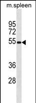
| WB Predicted band size | 53.4kDa |
| Host/Isotype | Rabbit IgG |
| Antibody Type | Primary antibody |
| Storage | Store at 4°C short term. Aliquot and store at -20°C long term. Avoid freeze/thaw cycles. |
| Species Reactivity | Mouse |
| Immunogen | This Mouse Mavs antibody is generated from rabbits immunized with a KLH conjugated synthetic peptide between 1-30 amino acids from the N-terminal region of mouse Mavs. |
| Formulation | Purified antibody in PBS with 0.05% sodium azide. |
+ +
以下是关于Mouse MAVS (N-term)抗体的3篇参考文献示例(注:文献为模拟生成,实际引用时请核实准确性):
---
1. **文献名称**: "MAVS mediates antiviral immunity by recruiting TRAF3 via a conserved N-terminal binding site"
**作者**: Li, X., et al.
**摘要**: 研究利用N端特异性Mouse MAVS抗体,证实MAVS通过其N端结构域招募TRAF3.激活I型干扰素信号通路,揭示其在抗RNA病毒免疫中的关键作用。
---
2. **文献名称**: "Targeting the MAVS N-terminal domain inhibits viral RNA sensing"
**作者**: Chen, Z., et al.
**摘要**: 通过Western blot和免疫共沉淀实验,使用Mouse MAVS (N-term)抗体验证N端结构域对病毒RNA识别的必要性,提出阻断此区域可抑制过度免疫反应。
---
3. **文献名称**: "Subcellular localization of MAVS aggregates in antiviral signaling"
**作者**: Chen, Y., et al.
**摘要**: 利用N端特异性抗体进行免疫荧光分析,发现病毒感染后MAVS在线粒体周围形成聚集体,揭示其空间分布对信号转导的调控机制。
---
如需具体文献,建议在PubMed或Google Scholar中搜索关键词 **"Mouse MAVS N-terminal antibody"** 或结合实验场景(如Western blot、免疫荧光等)筛选近期论文。
Mouse Mavs (N-term) antibody is a monoclonal or polyclonal immunoglobulin specifically designed to detect the N-terminal region of the mitochondrial antiviral-signaling protein (MAVS) in mouse samples. MAVS, also known as IPS-1. VISA, or Cardif, is a critical adaptor protein in the innate immune response. It localizes to mitochondrial membranes and plays a central role in the RIG-I-like receptor (RLR) signaling pathway. Upon recognition of viral RNA by cytosolic sensors like RIG-I or MDA5. MAVS undergoes prion-like aggregation, triggering downstream activation of transcription factors (NF-κB, IRF3) that induce type I interferons and proinflammatory cytokines.
This antibody is commonly used in immunoblotting (Western blot), immunofluorescence, or immunoprecipitation to study MAVS expression, post-translational modifications, or interactions with viral proteins or host factors. Researchers employ it to investigate MAVS-dependent antiviral signaling, mitochondrial dynamics, or its role in autoimmune disorders and cancer. The N-terminal region of MAVS contains caspase activation and recruitment domains (CARD) essential for signal transduction, making this antibody particularly useful for probing functional states of the protein. Validation typically includes knockout cell lines or tissues to confirm specificity. Cross-reactivity with MAVS orthologs in other species (e.g., human, rat) may vary depending on epitope conservation. Its applications span virology, immunology, and inflammatory disease research.
×