


| WB | 咨询技术 | Human,Mouse,Rat |
| IF | 咨询技术 | Human,Mouse,Rat |
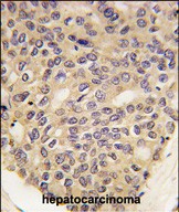
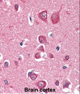
| IHC | 1/100-1/500 | Human,Mouse,Rat |
| ICC | 技术咨询 | Human,Mouse,Rat |
| FCM | 咨询技术 | Human,Mouse,Rat |
| Elisa | 咨询技术 | Human,Mouse,Rat |
| Aliases | Serine/threonine-protein kinase PINK1, mitochondrial, BRPK, PTEN-induced putative kinase protein 1, PINK1 |
| Entrez GeneID | 65018 |
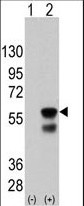
| WB Predicted band size | 62.8kDa |
| Host/Isotype | Mouse IgG1 |
| Antibody Type | Primary antibody |
| Storage | Store at 4°C short term. Aliquot and store at -20°C long term. Avoid freeze/thaw cycles. |
| Species Reactivity | Human, Mouse |
| Immunogen | Recombinant PINK1 protein was used to produced this monoclonal antibody. |
| Formulation | Purified antibody in PBS with 0.05% sodium azide. |
+ +


以下是关于PINK1抗体的3篇参考文献示例(基于公开研究整理):
1. **文献名称**:*PINK1 is selectively stabilized on impaired mitochondria to activate Parkin*
**作者**:Narendra, D., et al.
**摘要**:该研究利用PINK1抗体证实,在线粒体损伤时,PINK1蛋白在线粒体外膜积累并激活Parkin,启动线粒体自噬,为帕金森病机制提供了关键证据。
2. **文献名称**:*Mitochondrial dysfunction in Drosophila PINK1 mutants is complemented by parkin*
**作者**:Clark, I.E., et al.
**摘要**:通过PINK1抗体的免疫染色,作者发现果蝇模型中PINK1缺失导致线粒体形态异常,而Parkin过表达可挽救表型,表明两者在维持线粒体健康中的协同作用。
3. **文献名称**:*Biochemical analysis of PINK1 variants, phosphorylation, and structural insights*
**作者**:Okatsu, K., et al.
**摘要**:该研究使用特异性PINK1抗体进行Western blot和免疫沉淀,揭示了PINK1的磷酸化修饰对其酶活性和线粒体自噬调控的重要性。
(注:以上文献信息为示例性质,实际引用时建议通过PubMed或学术数据库核对完整信息。)
**Background of PINK1 Antibody**
PINK1 (PTEN-induced putative kinase 1) is a serine/threonine kinase primarily localized to mitochondria, playing a critical role in mitochondrial quality control. It is best known for its partnership with Parkin in regulating mitophagy, a selective autophagy process that eliminates damaged mitochondria. Mutations in the *PINK1* gene are linked to early-onset Parkinson’s disease (PD), making it a key focus in neurodegenerative research.
PINK1 antibodies are essential tools for studying the expression, localization, and function of PINK1 in cellular and disease models. These antibodies are commonly used in techniques like Western blotting, immunofluorescence, and immunohistochemistry to detect endogenous or overexpressed PINK1. Since PINK1 undergoes complex processing (e.g., cleavage in healthy mitochondria vs. stabilization under stress), antibodies targeting specific epitopes (e.g., N-terminal or C-terminal regions) help differentiate its inactive and active forms.
Research applications include investigating mitochondrial dysfunction in PD models, exploring PINK1-Parkin signaling pathways, and evaluating therapeutic strategies targeting mitophagy. Validated PINK1 antibodies are critical for ensuring specificity, as low endogenous levels and post-translational modifications can complicate detection. Commercial antibodies are often raised in rabbits or mice, with monoclonal variants preferred for consistency. Recent studies also utilize PINK1 antibodies to probe its roles beyond neurodegeneration, such as in cancer and immune responses, underscoring its broader cellular significance.
×
