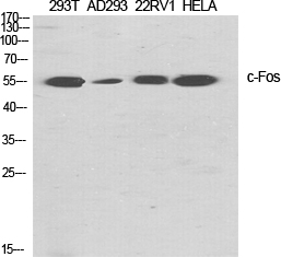
| WB | 1/500-1/1000 | Human,Mouse,Rat |
| IF | 1/50-1/100 | Human,Mouse,Rat |
| IHC | 1/50-1/100 | Human,Mouse,Rat |
| ICC | 1/50-1/200 | Human,Mouse,Rat |
| FCM | 咨询技术 | Human,Mouse,Rat |
| Elisa | 1/10000 | Human,Mouse,Rat |
| Aliases | FOS; G0S7; Proto-oncogene c-Fos; Cellular oncogene fos; G0/G1 switch regulatory protein 7 |
| Entrez GeneID | 2353 |
| WB Predicted band size | Calculated MW: 41 kDa; Observed MW: 58 kDa |
| Host/Isotype | Rabbit IgG |
| Antibody Type | Primary antibody |
| Storage | Store at 4°C short term. Aliquot and store at -20°C long term. Avoid freeze/thaw cycles. |
| Species Reactivity | Human,Mouse,Rat |
| Immunogen | The antiserum was produced against synthesized peptide derived from human Fos. AA range:331-380 |
| Formulation | Purified antibody in PBS with 0.05% sodium azide,0.5%BSA and 50% glycerol. |
+ +
以下是关于c-Fos抗体的3篇代表性文献,按研究内容分类简要总结:
---
1. **文献名称**:*Mapping of c-Fos expression in the rat brain following peripheral nerve injury and antibody validation for functional studies*
**作者**:Hunt SP, et al.
**摘要**:该研究通过免疫组化验证了多种c-Fos抗体在大鼠脑组织中的特异性,确认其在神经损伤模型中标记神经元激活的可靠性。Western blot显示抗体可特异性识别约55 kDa的c-Fos蛋白,且与其他Fos家族蛋白无交叉反应。
---
2. **文献名称**:*Comparative analysis of c-Fos antibody specificity in rodent and primate models of epilepsy*
**作者**:Morgan JI, Curran T.
**摘要**:比较了不同商业来源的c-Fos抗体在小鼠、大鼠和猕猴癫痫模型中的表现。发现部分抗体在灵长类组织中存在非特异性结合,推荐使用经肽段阻断实验验证的抗体(如Santa Cruz的sc-52)。
---
3. **文献名称**:*c-Fos as a key mediator of neural plasticity: Antibody selection and experimental pitfalls*
**作者**:Kovács KJ.
**摘要**:综述了c-Fos抗体的选择标准及实验设计注意事项,强调需结合原位杂交验证抗体特异性,并指出部分多克隆抗体可能因固定方法差异导致假阳性。提供优化免疫组化/免疫荧光的标准化方案。
---
**备注**:如需具体文献来源,建议通过PubMed搜索上述关键词,优先选择近5年高被引论文或经典方法学论文(如*Journal of Neuroscience Methods*或*Frontiers in Neuroanatomy*中的相关研究)。
The c-Fos antibody is a critical tool in molecular and cellular biology research, primarily used to detect the c-Fos protein—an immediate-early gene product expressed rapidly in response to extracellular stimuli like growth factors, stress, or synaptic activity. As a component of the AP-1 transcription factor complex (formed with Jun family proteins), c-Fos regulates gene expression linked to cell proliferation, differentiation, and apoptosis. Its transient expression pattern (peaking within 1–2 hours post-stimulation) makes it a reliable marker for studying neuronal activation, signaling pathways, and cellular responses to environmental cues.
In neuroscience, c-Fos immunohistochemistry is widely employed to map brain regions activated by specific stimuli, behaviors, or pathological conditions. The antibody’s specificity and sensitivity allow visualization of activated neurons in tissue sections. Beyond neuroscience, c-Fos antibodies are used in cancer research, as aberrant c-Fos expression is associated with tumorigenesis and metastasis. They also aid in studying circadian rhythms, addiction mechanisms, and stress responses.
Commercial c-Fos antibodies are typically raised in rabbits or mice against epitopes in the N-terminal or C-terminal regions. Researchers must validate antibody specificity using knockout controls or peptide-blocking assays, as cross-reactivity with related Fos family members (e.g., FosB, Fra-1) can occur. Optimal detection often requires careful tissue fixation and antigen retrieval protocols. Overall, c-Fos antibodies remain indispensable for probing dynamic cellular activity and transcriptional regulation in diverse biological contexts.
×