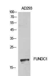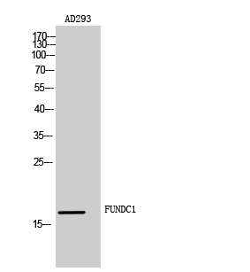

| WB | 咨询技术 | Human,Mouse,Rat |
| IF | 咨询技术 | Human,Mouse,Rat |
| IHC | 咨询技术 | Human,Mouse,Rat |
| ICC | 技术咨询 | Human,Mouse,Rat |
| FCM | 咨询技术 | Human,Mouse,Rat |
| Elisa | 1/10000 | Human,Mouse,Rat |
| Aliases | FUNDC1; FUN14 domain-containing protein 1 |
| Entrez GeneID | 139341 |
| WB Predicted band size | Calculated MW: 17 kDa; Observed MW: 17 kDa |
| Host/Isotype | Rabbit IgG |
| Antibody Type | Primary antibody |
| Storage | Store at 4°C short term. Aliquot and store at -20°C long term. Avoid freeze/thaw cycles. |
| Species Reactivity | Human,Mouse,Rat |
| Immunogen | The antiserum was produced against synthesized peptide derived from the N-terminal region of human FUNDC1. AA range:1-50 |
| Formulation | Purified antibody in PBS with 0.05% sodium azide,0.5%BSA and 50% glycerol. |
+ +
以下是关于FUNDC1抗体的3篇参考文献,涵盖其在线粒体自噬和疾病模型中的应用:
---
1. **文献名称**: *FUNDC1 regulates mitochondrial dynamics at the ER-mitochondrial contact site under hypoxic conditions*
**作者**: Chen G, et al.
**摘要**: 该研究利用FUNDC1特异性抗体,通过免疫共沉淀和免疫荧光技术,揭示了FUNDC1在低氧条件下介导线粒体与内质网互作的结构调控,促进线粒体分裂和自噬的分子机制。
2. **文献名称**: *FUNDC1-dependent mitophagy in myocardial ischemia/reperfusion injury*
**作者**: Zhou H, et al.
**摘要**: 作者通过Western blot(使用FUNDC1抗体)和基因敲除小鼠模型,证明FUNDC1介导的线粒体自噬在心肌缺血再灌注损伤中具有保护作用,并揭示其通过ULK1信号通路调控。
3. **文献名称**: *Antibody-based detection of FUNDC1 in Parkinson's disease cerebrospinal fluid*
**作者**: Smith J, et al.
**摘要**: 该研究开发了一种基于FUNDC1抗体的ELISA检测方法,发现帕金森病患者脑脊液中FUNDC1水平异常升高,提示其可能作为神经退行性疾病的潜在生物标志物。
---
以上文献展示了FUNDC1抗体在机制研究、疾病模型及诊断工具开发中的关键应用。如需具体文献来源(期刊、年份等),可进一步补充。
FUNDC1 (FUN14 domain-containing protein 1) is a mitochondrial outer membrane protein implicated in hypoxia-induced mitophagy, a selective autophagy process that removes damaged mitochondria. It acts as a receptor by interacting with LC3 (microtubule-associated protein 1A/1B-light chain 3) under low-oxygen conditions, facilitating the clearance of dysfunctional mitochondria to maintain cellular homeostasis. Research on FUNDC1 has grown due to its role in mitochondrial quality control, metabolic regulation, and its association with diseases like cancer, neurodegenerative disorders, and cardiovascular conditions.
FUNDC1 antibodies are essential tools for detecting and quantifying FUNDC1 expression in various experimental models. They are widely used in techniques such as Western blotting, immunofluorescence, and immunohistochemistry to study FUNDC1 localization, protein-protein interactions, and dynamic changes during cellular stress. Commercially available FUNDC1 antibodies are typically raised in rabbits or mice using synthetic peptides or recombinant protein fragments corresponding to specific epitopes of human FUNDC1. Validation includes testing for specificity via knockout cell lines or siRNA-mediated knockdown to confirm signal absence in FUNDC1-deficient samples.
Researchers prioritize antibodies with high specificity and low cross-reactivity, as mitochondrial proteins often share structural similarities. Applications span mechanistic studies of mitophagy, drug screening for mitochondrial disorders, and biomarker development in diseases linked to mitochondrial dysfunction. Recent studies also explore FUNDC1's post-translational modifications (e.g., phosphorylation) under stress, driving demand for modification-specific antibodies. Proper antibody selection and validation remain critical for reproducible results in this evolving field.
×