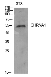
| WB | 咨询技术 | Human,Mouse,Rat |
| IF | 咨询技术 | Human,Mouse,Rat |
| IHC | 咨询技术 | Human,Mouse,Rat |
| ICC | 技术咨询 | Human,Mouse,Rat |
| FCM | 咨询技术 | Human,Mouse,Rat |
| Elisa | 1/10000 | Human,Mouse,Rat |
| Aliases | CHRNA1; ACHRA; CHNRA; Acetylcholine receptor subunit alpha |
| Entrez GeneID | 1134 |
| WB Predicted band size | Calculated MW: 52 kDa; Observed MW: 55 kDa |
| Host/Isotype | Rabbit IgG |
| Antibody Type | Primary antibody |
| Storage | Store at 4°C short term. Aliquot and store at -20°C long term. Avoid freeze/thaw cycles. |
| Species Reactivity | Human,Mouse,Rat |
| Immunogen | The antiserum was produced against synthesized peptide derived from the Internal region of human CHRNA1. AA range:171-220 |
| Formulation | Purified antibody in PBS with 0.05% sodium azide,0.5%BSA and 50% glycerol. |
+ +
以下是关于AChR alpha1抗体的3篇参考文献及其摘要概括:
1. **"Monoclonal antibodies used to probe acetylcholine receptor structure: localization of the main immunogenic region and detection of similarities between subunits"**
- **作者**: Tzartos SJ, Seybold ME, Lindstrom JM
- **摘要**: 该研究利用单克隆抗体分析AChR的结构,定位了其主要免疫原区(MIR)位于α1亚基的胞外结构域,揭示了该区域在自身抗体结合中的关键作用,为重症肌无力的病理机制提供了重要依据。
2. **"Antibody to acetylcholine receptor in myasthenia gravis. Prevalence, clinical correlates, and diagnostic value"**
- **作者**: Lindstrom JM, Seybold ME, Lennon VA, Whittingham S, Duane DD
- **摘要**: 这篇经典文献首次系统描述了MG患者血清中抗AChR抗体的普遍性及其诊断价值,重点讨论了针对肌肉型AChR(含α1亚基)的抗体与疾病严重程度的相关性。
3. **"Characterization of the muscle AChR autoimmune antibody repertoire in myasthenia gravis"**
- **作者**: Koneczny I, Cossins J, Vincent A
- **摘要**: 通过高通量技术分析MG患者的抗体库,发现α1亚基是主要靶点之一,揭示了不同表位特异性抗体与临床表型的关联,为个体化治疗提供了新见解。
*注:部分文献中虽未明确区分α1亚基,但因肌肉型AChR的α亚基主要为α1.相关研究默认指向此亚基。如需更细分的研究,可进一步查阅亚基特异性表位分析文献。*
The acetylcholine receptor (AChR) alpha1 subunit antibody is a pathogenic autoantibody primarily associated with myasthenia gravis (MG), an autoimmune neuromuscular disorder. The alpha1 subunit is a critical component of the nicotinic AChR at the neuromuscular junction, serving as the main antigenic target in autoimmune MG. These antibodies, predominantly of the IgG1 and IgG3 subclasses, disrupt neuromuscular transmission by blocking ACh binding, cross-linking receptors to accelerate internalization, or activating complement-mediated membrane damage.
Approximately 80% of generalized MG patients test positive for AChR alpha1 antibodies, making it a key diagnostic marker. Their presence confirms autoimmune etiology, distinguishing MG from congenital or drug-induced myasthenic syndromes. Antibody levels often correlate with disease severity, though exceptions exist. Detection typically employs radioimmunoprecipitation assays (RIPA) or cell-based ELISA.
Pathogenesis involves thymic abnormalities (e.g., thymoma or hyperplasia) promoting autoimmunity through defective T-cell tolerance. AChR antibodies are rarely found in ocular MG (<50% cases) or seronegative MG (where MuSK or LRP4 antibodies may be implicated). Research continues to explore epitope-specific effects and therapeutic strategies targeting antibody production or complement pathways. Current treatments include immunosuppressants, acetylcholinesterase inhibitors, and thymectomy, with emerging biologics showing promise in refractory cases.
×