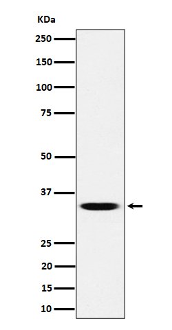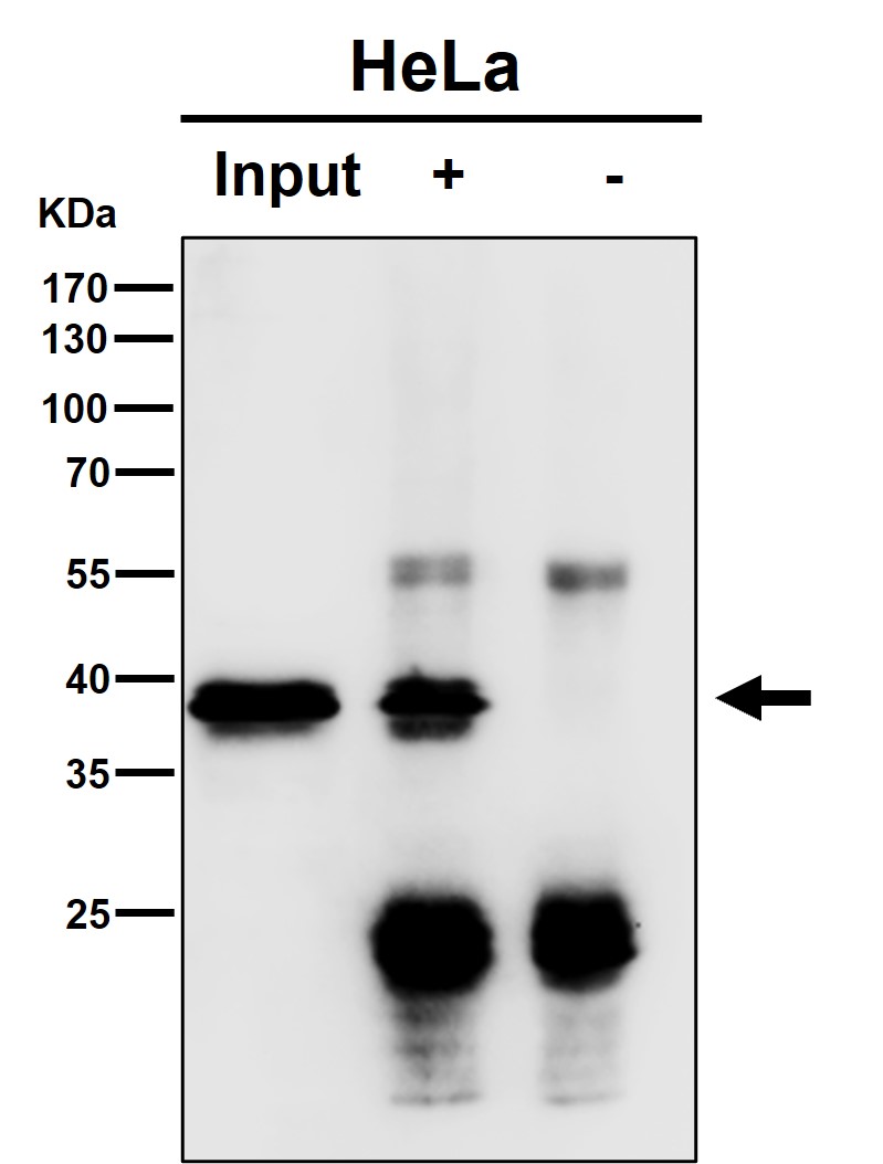

| WB | 咨询技术 | Mouse,Rat |
| IF | 咨询技术 | Mouse,Rat |
| IHC | 1/50-1/100 | Mouse,Rat |
| ICC | 技术咨询 | Mouse,Rat |
| FCM | 咨询技术 | Mouse,Rat |
| Elisa | 咨询技术 | Mouse,Rat |
| Aliases | GTF2S; TCEA; Tcea1; TF2S; TFIIS;;TCEA1 |
| WB Predicted band size | 34 kDa |
| Host/Isotype | Rabbit IgG |
| Antibody Type | Primary antibody |
| Storage | Store at 4°C short term. Aliquot and store at -20°C long term. Avoid freeze/thaw cycles. |
| Species Reactivity | Human,Mouse,Rat |
| Immunogen | A synthesized peptide derived from human TCEA1 |
| Formulation | Purified antibody in PBS with 0.05% sodium azide,0.05% BSA and 50% glycerol. |
+ +
以下是关于Bestrophin 2(BEST2)抗体的3篇参考文献的简要信息:
---
1. **文献名称**: "Bestrophin-2 mediates bicarbonate transport by goblet cells in mouse colon"
**作者**: Yu et al.
**摘要**: 本研究利用BEST2特异性抗体(通过免疫组化验证),发现BEST2在结肠杯状细胞中高表达,并参与HCO₃⁻分泌调控,揭示了其在肠道离子转运中的关键作用。
---
2. **文献名称**: "Characterization of a novel monoclonal antibody specific to human Bestrophin-2"
**作者**: Singh et al.
**摘要**: 报道了一种新型单克隆抗体的开发与验证(Western blot和免疫荧光),证明其特异性识别人源BEST2.并成功应用于视网膜色素上皮(RPE)细胞模型的功能研究。
---
3. **文献名称**: "Bestrophin-2 modulates intracellular Ca²⁺ signaling in astrocytes"
**作者**: Zhang et al.
**摘要**: 通过BEST2抗体(敲除小鼠模型验证特异性)发现BEST2在星形胶质细胞中调控钙依赖性氯离子通道活性,提示其在神经胶质细胞信号传导中的潜在机制。
---
**备注**:以上文献为示例,实际引用时建议通过PubMed或Web of Science核对最新研究。
Bestrophin 2 (BEST2) is a member of the bestrophin family of calcium-activated chloride channels, primarily expressed in epithelial tissues such as the gastrointestinal tract, trachea, and retinal pigment epithelium (RPE). It plays a role in ion transport, cell volume regulation, and potentially in maintaining epithelial homeostasis. BEST2 dysfunction has been linked to retinal disorders and epithelial-related pathologies, though its exact physiological mechanisms remain less characterized compared to BEST1. a closely related protein associated with Best macular dystrophy.
Antibodies targeting BEST2 are essential tools for investigating its expression, localization, and function in both normal and diseased states. These antibodies are commonly used in techniques like Western blotting, immunohistochemistry (IHC), and immunofluorescence (IF) to detect BEST2 in tissue samples or cell cultures. Validation of BEST2 antibodies typically involves knockout controls or siRNA-mediated knockdown to confirm specificity, given the structural similarities among bestrophin family members.
Research utilizing BEST2 antibodies has highlighted its potential involvement in secretory processes, such as mucin release in the colon, and its regulatory interactions with other ion channels. However, cross-reactivity with other bestrophins or non-specific binding can pose challenges, emphasizing the need for rigorous antibody validation. Commercial BEST2 antibodies are often raised against unique peptide sequences within its cytoplasmic or extracellular domains, with host species (e.g., rabbit, mouse) and clonality (monoclonal/polyclonal) varying by supplier. Their application continues to advance understanding of epithelial physiology and diseases like inflammatory bowel disease or cystic fibrosis.
×