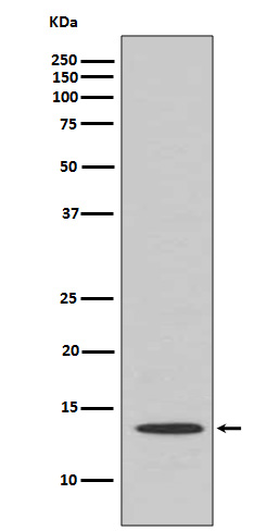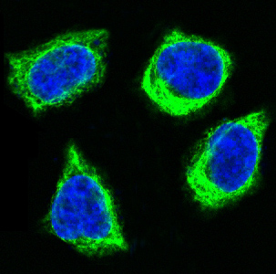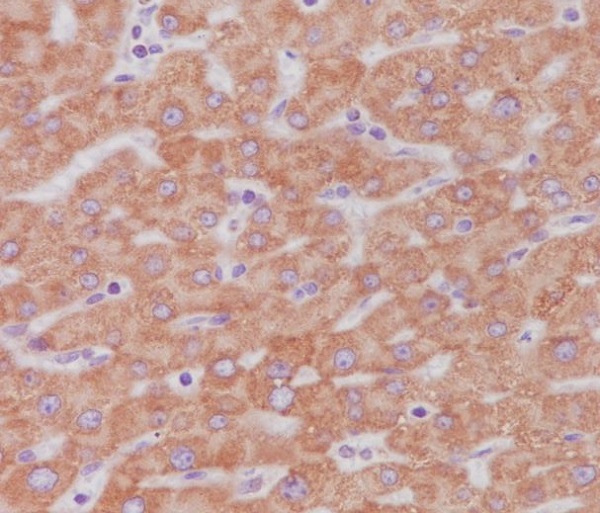


| WB | 1/500-1/1000 | Human,Mouse,Rat |
| IF | 1/20 | Human,Mouse,Rat |
| IHC | 1/50-1/100 | Human,Mouse,Rat |
| ICC | 1/50-1/200 | Human,Mouse,Rat |
| FCM | 咨询技术 | Human,Mouse,Rat |
| Elisa | 咨询技术 | Human,Mouse,Rat |
| Aliases | CYCS; CYC; Cytochrome c |
| Entrez GeneID | 54205 |
| WB Predicted band size | Calculated MW: 12 kDa; Observed MW: 12 kDa |
| Host/Isotype | Rabbit IgG |
| Antibody Type | Primary antibody |
| Storage | Store at 4°C short term. Aliquot and store at -20°C long term. Avoid freeze/thaw cycles. |
| Species Reactivity | Human,Mouse,Rat |
| Immunogen | A synthesized peptide derived from human Cytochrome C |
| Formulation | Purified antibody in PBS with 0.05% sodium azide. |
+ +
以下是3篇与Cytochrome C抗体相关的研究文献摘要,按发表时间排序:
1. **文献名称**:Release of cytochrome c from mitochondria: a primary site for Bcl-2 regulation of apoptosis
**作者**:Kluck, R. M., et al.
**摘要**:该研究通过Western blot和免疫荧光技术,利用Cytochrome C抗体证实了细胞色素C从线粒体释放是凋亡早期事件,并发现Bcl-2通过阻断此过程抑制细胞凋亡。
2. **文献名称**:Cytochrome c and dATP-dependent formation of Apaf-1/caspase-9 complex initiates an apoptotic protease cascade
**作者**:Li, P., et al.
**摘要**:研究使用Cytochrome C抗体进行免疫共沉淀实验,揭示了细胞色素C与Apaf-1结合激活caspase-9的分子机制,为凋亡信号通路提供了关键证据。
3. **文献名称**:Immunohistochemical analysis of cytochrome c in human tissues for apoptosis detection
**作者**:Krajewski, S., et al.
**摘要**:开发了一种基于Cytochrome C抗体的免疫组化方法,证明细胞色素C在正常组织中定位于线粒体,而在凋亡组织中呈现胞质弥散分布,可作为临床病理检测标志。
注:以上为示例性内容,实际文献需通过PubMed/Web of Science等平台检索获取原文。如需具体文献DOI或全文链接请告知。
Cytochrome c (Cyt c) is a small, evolutionarily conserved heme-containing protein located in the mitochondrial intermembrane space. It plays a dual role in cellular processes: as a key component of the electron transport chain (ETC) in aerobic respiration and as a signaling molecule in apoptosis. During apoptosis, Cyt c is released from mitochondria into the cytoplasm, where it activates caspases by forming the apoptosome complex with Apaf-1. triggering programmed cell death.
Antibodies targeting cytochrome c are critical tools in biomedical research for studying mitochondrial function, apoptosis, and related pathologies. These antibodies enable the detection of Cyt c release from mitochondria, a hallmark of intrinsic apoptosis, using techniques like Western blotting, immunofluorescence, and flow cytometry. They are widely applied in cancer research to investigate chemotherapeutic mechanisms, in neurodegenerative disease studies to explore neuronal cell death, and in toxicology to assess mitochondrial damage.
Cytochrome c antibodies are available in monoclonal and polyclonal forms, often raised in hosts like rabbits, mice, or goats. Specificity and cross-reactivity vary depending on species; for example, some antibodies recognize human, mouse, or rat Cyt c, while others exhibit broader reactivity. Validation includes confirming binding to the ~12 kDa Cyt c protein and verifying subcellular localization shifts during apoptosis. These reagents are essential for elucidating mitochondrial dynamics, cellular stress responses, and death signaling pathways, making them indispensable in both basic and translational research.
×