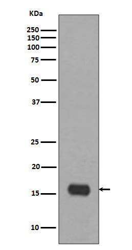


| WB | 1/500-1/1000 | Human,Mouse,Rat |
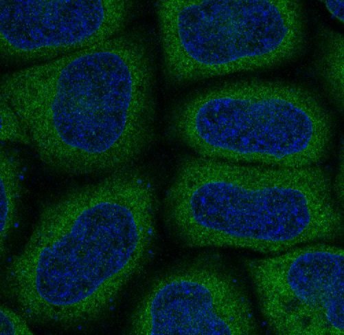
| IF | 1/20 | Human,Mouse,Rat |
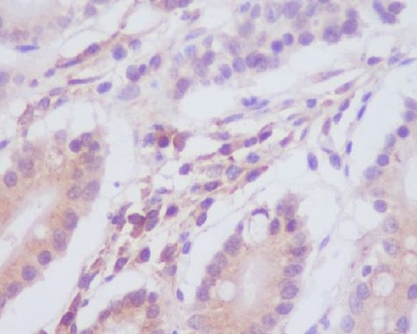
| IHC | 1/50-1/100 | Human,Mouse,Rat |
| ICC | 1/50-1/200 | Human,Mouse,Rat |
| FCM | 1/50-1/100 | Human,Mouse,Rat |
| Elisa | 咨询技术 | Human,Mouse,Rat |
| Aliases | Microtubule-associated proteins 1A/1B light chain 3A; Autophagy-related protein LC3 A; Autophagy-related ubiquitin-like modifier LC3 A; MAP1 light chain 3-like protein 1; MAP1A/MAP1B light chain 3 A; MAP1A/MAP1B LC3 A; Microtubule-associated protein 1 light chain 3 alpha |
| Entrez GeneID | 84557 |
| WB Predicted band size | Calculated MW: 14 kDa; Observed MW: 14 kDa |
| Host/Isotype | Rabbit IgG |
| Antibody Type | Primary antibody |
| Storage | Store at 4°C short term. Aliquot and store at -20°C long term. Avoid freeze/thaw cycles. |
| Species Reactivity | Human,Mouse,Rat |
| Immunogen | A synthesized peptide derived from human MAP1LC3A |
| Formulation | Purified antibody in PBS with 0.05% sodium azide. |
+ +
以下是关于LC3A抗体的3篇参考文献及其摘要概括:
---
1. **文献名称**:*Guidelines for the use and interpretation of assays for monitoring autophagy*
**作者**:Klionsky, D.J. et al.
**摘要**:该指南系统总结了自噬研究中的常用方法,包括LC3A抗体的应用。文中强调LC3A作为自噬体标记物的特异性,并讨论了其在Western blot和免疫荧光中的使用注意事项,如需结合溶酶体抑制剂以准确检测自噬通量。
---
2. **文献名称**:*LC3A-positive light microscopy detected patterns of autophagy and prognosis in operable breast carcinomas*
**作者**:Sivridis, E. et al.
**摘要**:研究通过LC3A抗体进行免疫组化分析,发现乳腺癌组织中特定的自噬相关结构(如“stone-like”结构)与患者较差预后相关。该文献支持LC3A抗体在肿瘤自噬临床评估中的潜在价值。
---
3. **文献名称**:*Differential expression patterns of LC3A and LC3B in cancer: A tissue microarray analysis*
**作者**:Karpathiou, G. et al.
**摘要**:利用组织微阵列技术比较LC3A和LC3B在多种癌症中的表达差异。研究发现LC3A在侵袭性肿瘤中高表达,且其抗体特异性在区分不同自噬阶段中表现优于LC3B,提示亚型特异性抗体的重要性。
---
**备注**:以上文献可从PubMed或Web of Science通过标题/作者检索获取全文。若需实验细节,推荐优先查阅Klionsky团队的自噬指南(定期更新版)。
The LC3A antibody is a crucial tool for studying autophagy, a cellular degradation process. LC3 (Microtubule-associated protein 1A/1B-light chain 3) belongs to the ATG8 protein family, with three mammalian isoforms: LC3A, LC3B, and LC3C. LC3A exists as two splice variants (LC3A-a and LC3A-b) differing in their C-terminal regions. During autophagy, cytosolic LC3-I is conjugated to phosphatidylethanolamine, forming membrane-bound LC3-II, which integrates into autophagosome membranes. LC3A antibodies specifically detect these lipidated and non-lipidated forms, enabling autophagy flux assessment via immunoblotting, immunofluorescence, or immunohistochemistry.
While LC3B is widely expressed, LC3A shows tissue-specific patterns, with higher expression in epithelial cells and certain tumors. Its role extends beyond canonical autophagy, potentially involving inflammation, cancer progression, and pathogen clearance. Researchers use LC3A antibodies to differentiate isoform-specific autophagy mechanisms or pathological contexts. However, cross-reactivity with other LC3 isoforms requires validation via knockout controls. Commercial antibodies often target conserved N-terminal regions but may vary in specificity. LC3A puncta formation observed under microscopy serves as a qualitative marker for autophagosomes, while quantitative methods (e.g., LC3-II/I ratio) reflect autophagic activity. Its applications span cancer research, neurodegenerative disease studies, and investigations of infectious host-pathogen interactions.
×