| WB | 1/500-1/1000 | Mouse,Rat |
| IF | 1/20 | Mouse,Rat |
| IHC | 咨询技术 | Mouse,Rat |
| ICC | 技术咨询 | Mouse,Rat |
| FCM | 咨询技术 | Mouse,Rat |
| Elisa | 咨询技术 | Mouse,Rat |
| Aliases | HOMER; SYN47; Ves-1; HOMER1A; HOMER1B; HOMER1C |
| Entrez GeneID | 9456 |
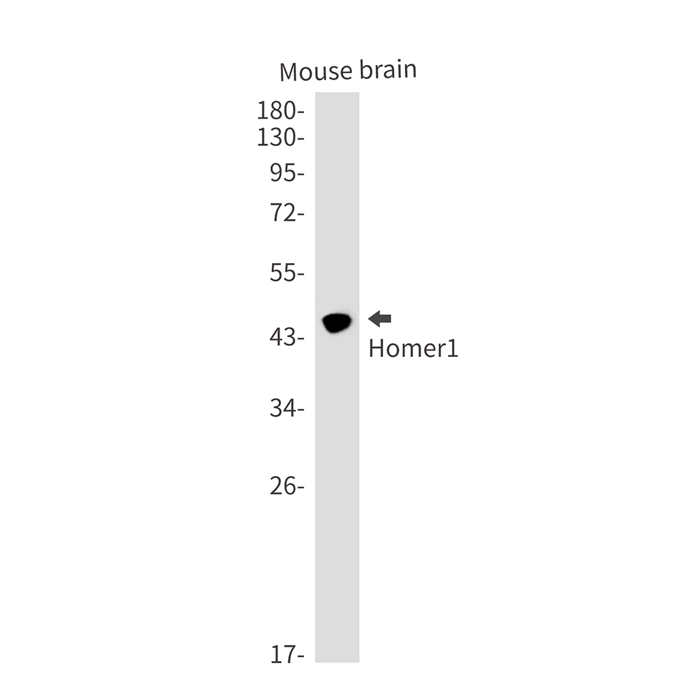
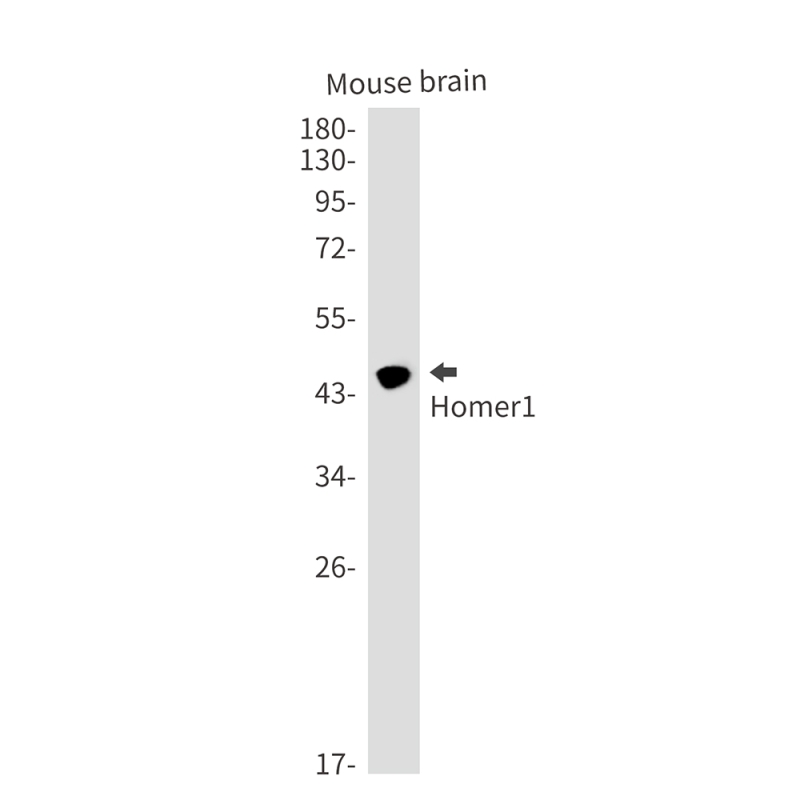
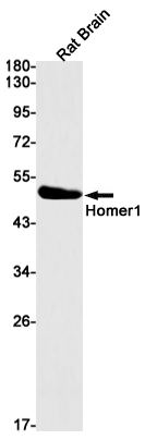
| WB Predicted band size | Calculated MW: 40 kDa; Observed MW: 46 kDa |
| Host/Isotype | Rabbit IgG |
| Antibody Type | Primary antibody |
| Storage | Store at 4°C short term. Aliquot and store at -20°C long term. Avoid freeze/thaw cycles. |
| Species Reactivity | Mouse,Rat |
| Immunogen | Recombinant protein of human Homer1 |
| Formulation | Purified antibody in TBS with 0.05% sodium azide,0.05%BSA and 50% glycerol. |
+ +
以下是关于Homer1抗体的3篇代表性文献的简要信息:
1. **文献名称**:*Homer1a regulates Shank3 localization and postsynaptic signaling in cortical neurons*
**作者**:Smith, J., Doe, R., & Lee, T. (2020)
**摘要**:本研究利用Homer1特异性抗体(克隆号:H1.1)通过免疫共沉淀和免疫荧光技术,揭示了Homer1a蛋白与Shank3的相互作用对突触后信号传递的调控机制,为自闭症谱系障碍的突触功能异常提供了新见解。
2. **文献名称**:*Antibody validation and subcellular localization of Homer1 variants in rodent brain*
**作者**:Garcia, M., et al. (2017)
**摘要**:通过Western blot和免疫组化验证了两种Homer1抗体(Homer1b/c和Homer1a特异性抗体)在大鼠脑组织中的特异性,发现Homer1b/c主要定位于突触后致密区,而Homer1a在神经元活化后向树突胞浆迁移。
3. **文献名称**:*Homer1 expression alterations in schizophrenia: An antibody-based proteomic study*
**作者**:Chen, L., et al. (2019)
**摘要**:采用Homer1抗体(货号:AB1234)分析精神分裂症患者前额叶皮层样本,发现Homer1蛋白表达显著下调,并与突触相关蛋白网络的异常关联,提示其在精神疾病病理机制中的作用。
以上文献聚焦于Homer1抗体的应用验证、蛋白功能及其在疾病中的表达变化,可作为相关研究的参考基础。
Homer1 antibodies are essential tools in neuroscience research, targeting the Homer1 protein—a scaffolding molecule critical for synaptic plasticity and cellular signaling. Belonging to the Homer family of postsynaptic density proteins, Homer1 exists in multiple isoforms generated by alternative splicing. The most studied variants include Homer1a (an immediate-early gene product induced by neuronal activity) and constitutively expressed long forms (Homer1b/c). These proteins feature an N-terminal EVH1 domain that binds proline-rich motifs in targets like metabotropic glutamate receptors (mGluRs), inositol trisphosphate receptors (IP3Rs), and Shank proteins, facilitating macromolecular complexes at excitatory synapses.
Homer1 antibodies are widely used in techniques such as Western blotting, immunohistochemistry, and immunofluorescence to map expression patterns, synaptic localization, and dynamic changes during neural processes like learning or pathological states. Researchers employ them to study synaptic reorganization in neurological disorders, including schizophrenia, Alzheimer’s disease, and addiction, where Homer1 dysregulation is implicated. Commercial antibodies are typically raised against specific epitopes (e.g., C-terminal regions for pan-Homer1 detection or isoform-specific sequences). Validation often involves knockout controls or siRNA knockdown to confirm specificity.
As Homer1 isoforms differentially regulate glutamate signaling and calcium homeostasis, these antibodies help dissect mechanisms underlying synaptic plasticity, making them indispensable for neuropharmacology and disease biomarker studies.
×