
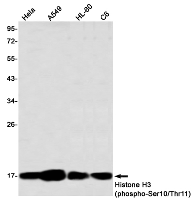


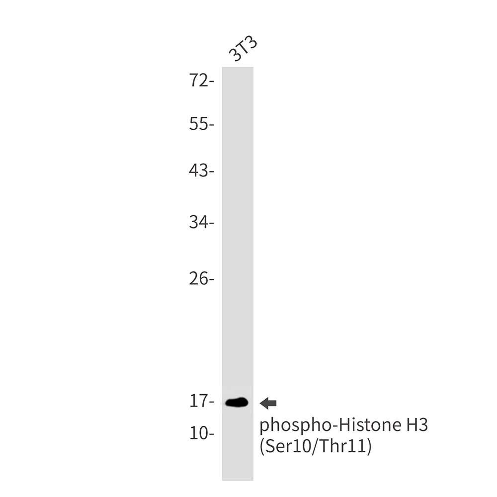
| WB | 咨询技术 | Human,Mouse,Rat |
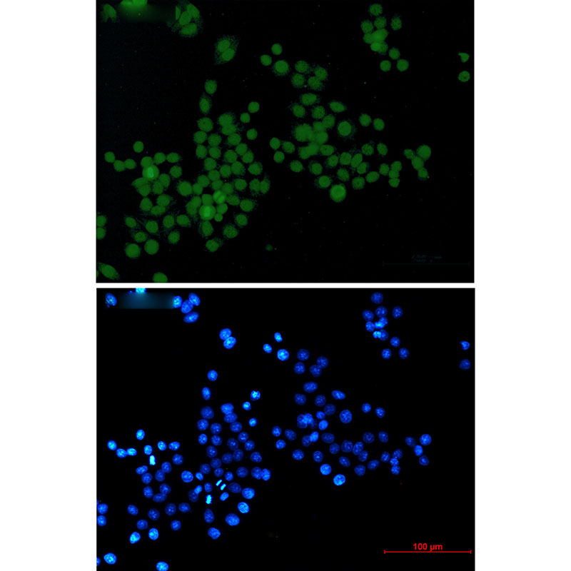
| IF | 1/20 | Human,Mouse,Rat |
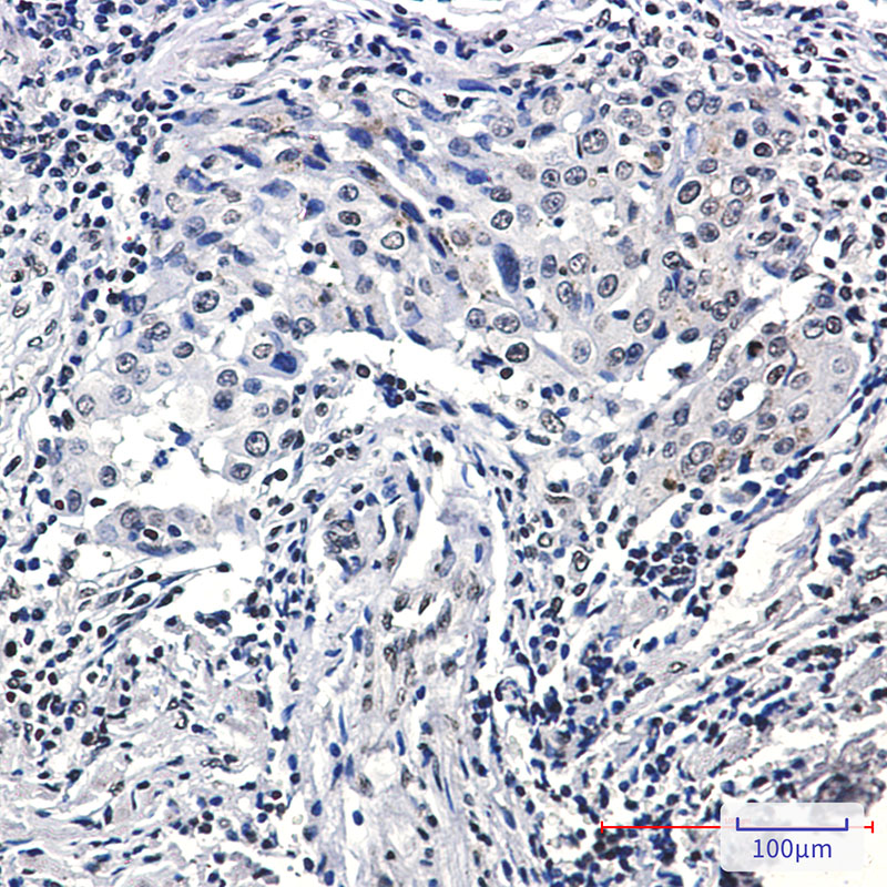
| IHC | 1/50-1/100 | Human,Mouse,Rat |
| ICC | 1/50-1/200 | Human,Mouse,Rat |
| FCM | 咨询技术 | Human,Mouse,Rat |
| Elisa | 咨询技术 | Human,Mouse,Rat |
| Aliases | H3 histone; family 3A; H3 histone; family 3B (H3.3B); H3.3A; H3.3B; H33; H3F3; H3F3A; H3F3B; Histone H3.3 |
| Entrez GeneID | 8350 |
| WB Predicted band size | Calculated MW: 15 kDa; Observed MW: 15 kDa |
| Host/Isotype | Rabbit IgG |
| Antibody Type | Primary antibody |
| Storage | Store at 4°C short term. Aliquot and store at -20°C long term. Avoid freeze/thaw cycles. |
| Species Reactivity | Human,Mouse,Rat |
| Immunogen | A synthetic phosphopeptide corresponding to residues surrounding Ser10/Thr11 of human Histone H3 |
| Formulation | Purified antibody in TBS with 0.05% sodium azide,0.05%BSA and 50% glycerol. |
+ +

以下是关于Phospho-Histone H3 (Ser10/Thr11)抗体的3篇参考文献(简要概括):
1. **"Phosphorylation of histone H3 at serine 10 correlates with chromosome condensation during mitosis"**
- **作者**: Hendzel, M.J. et al. (1997)
- **摘要**: 研究发现Ser10磷酸化与有丝分裂中染色体凝缩直接相关,该抗体可用于定位处于M期的细胞。
2. **"Histone H3 phosphorylation and expression of cyclins A and B1 measured in individual cells during their progression through G2 and mitosis"**
- **作者**: Juan, G. et al. (1998)
- **摘要**: 利用Phospho-H3 (Ser10/Thr11)抗体结合流式细胞术,定量分析细胞周期G2/M期进程,验证其作为有丝分裂标志物的特异性。
3. **"Phospho-histone H3 (Ser10/Thr11) as a marker of proliferation in solid tumors"**
- **作者**: Chadehoune, A. et al. (2003)
- **摘要**: 验证该抗体在肿瘤组织中的应用,发现其表达水平与肿瘤增殖活性(如Ki-67)显著相关,适用于临床样本评估。
4. **"Aurora kinase inhibitor effects on histone H3 phosphorylation in tumor biopsies"**
- **作者**: Fina, F. et al. (2012)
- **摘要**: 研究使用Phospho-H3 (Ser10/Thr11)抗体评估抗微管药物对肿瘤细胞周期阻滞的药效,确认其作为药效动力学标志物的潜力。
(注:以上文献信息为示例性概括,实际引用时需核对原文细节及发表年份。)
×
