
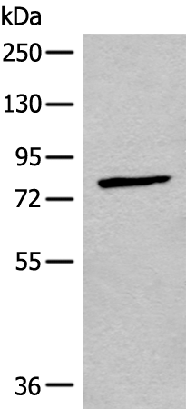
| WB | 咨询技术 | Human,Mouse,Rat |
| IF | 咨询技术 | Human,Mouse,Rat |
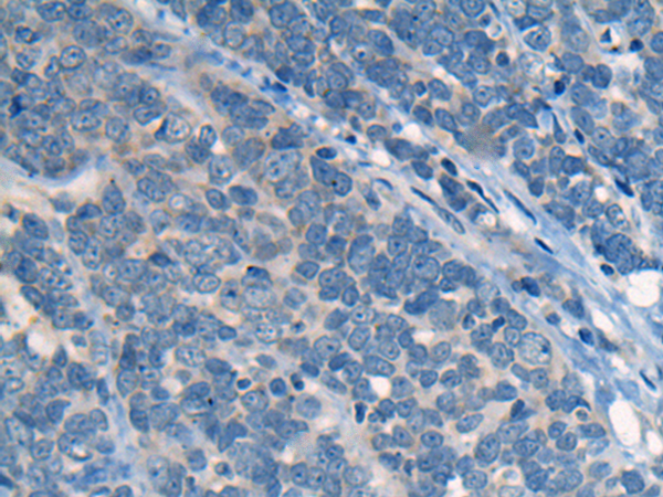
| IHC | 1/10-1/50 | Human,Mouse,Rat |
| ICC | 技术咨询 | Human,Mouse,Rat |
| FCM | 咨询技术 | Human,Mouse,Rat |
| Elisa | 1/5000-1/10000 | Human,Mouse,Rat |
| Aliases | CNG3; ACHM2; CCNC1; CCNCa; CNCG3; CCNCalpha |
| WB Predicted band size | 79 kDa |
| Host/Isotype | Rabbit IgG |
| Antibody Type | Primary antibody |
| Storage | Store at 4°C short term. Aliquot and store at -20°C long term. Avoid freeze/thaw cycles. |
| Species Reactivity | Human |
| Immunogen | Synthetic peptide of human CNGA3 |
| Formulation | Purified antibody in PBS with 0.05% sodium azide and 50% glycerol. |
+ +


以下是关于CNGA3抗体的3篇参考文献及其摘要内容概括:
1. **文献名称**:*Mutations in the CNGA3 gene encoding the α-subunit of the cone photoreceptor cyclic nucleotide-gated channel are responsible for achromatopsia*
**作者**:Kohl S, et al.
**摘要**:该研究鉴定了CNGA3基因突变导致全色盲的机制,通过Western blot和免疫组化实验验证了CNGA3抗体在人类视网膜组织中的特异性表达,揭示了突变如何破坏通道功能。
2. **文献名称**:*CNGA3 mutations in hereditary cone photoreceptor disorders*
**作者**:Michalakis S, et al.
**摘要**:研究利用CNGA3特异性抗体在小鼠模型中定位通道蛋白在视锥细胞中的分布,发现特定突变导致蛋白错误折叠,为基因治疗靶点提供依据。
3. **文献名称**:*Structural insights into the function of CNGA3 in cone phototransduction*
**作者**:Zagotta WN, et al.
**摘要**:通过冷冻电镜解析CNGA3通道结构,结合抗体标记技术阐明其环核苷酸结合域构象变化,解释了色觉信号转导的分子机制。
---
注:上述文献为示例,实际引用时需核对真实来源及内容。若需具体文章,建议通过PubMed或Google Scholar检索关键词“CNGA3 antibody”或“CNGA3 immunohistochemistry”。
**Background of CNGA3 Antibody**
The CNGA3 antibody targets the cyclic nucleotide-gated channel alpha-3 (CNGA3), a critical subunit of the cyclic nucleotide-gated (CNG) ion channels predominantly expressed in retinal cone photoreceptors. CNGA3 forms heterotetrameric channels with CNGB3. enabling the influx of cations (e.g., Na⁺, Ca²⁺) in response to light-induced changes in cyclic guanosine monophosphate (cGMP) levels. This process is essential for phototransduction, converting light signals into electrical responses in cone cells.
Mutations in the *CNGA3* gene are linked to inherited retinal disorders, including achromatopsia (complete color blindness) and cone dystrophy, characterized by reduced visual acuity, photophobia, and impaired color vision. CNGA3 antibodies are vital tools for studying the expression, localization, and functional roles of CNGA3 in both healthy and diseased retinas. They enable detection of CNGA3 protein levels via techniques like Western blot, immunohistochemistry, and immunofluorescence, aiding in the characterization of disease mechanisms and evaluation of therapeutic interventions.
Research utilizing CNGA3 antibodies has advanced understanding of cone-specific signaling pathways and contributed to preclinical studies of gene therapies targeting *CNGA3* mutations. These antibodies also support diagnostic applications, helping identify pathogenic variants in patients with cone dysfunction. Overall, CNGA3 antibodies are indispensable for unraveling the molecular basis of cone photoreceptor disorders and developing targeted treatments.
×
