


| WB | 咨询技术 | Human,Mouse,Rat |
| IF | 咨询技术 | Human,Mouse,Rat |
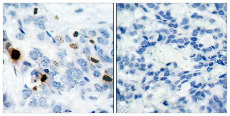
| IHC | 1/50-1/100 | Human,Mouse,Rat |
| ICC | 1/100-1/200 | Human,Mouse,Rat |
| FCM | 咨询技术 | Human,Mouse,Rat |
| Elisa | 咨询技术 | Human,Mouse,Rat |
| Aliases | H3/b, H3FB |
| Entrez GeneID | 8350;8351;8352;8353;8354;8355;8356;8357;8358;8968; |
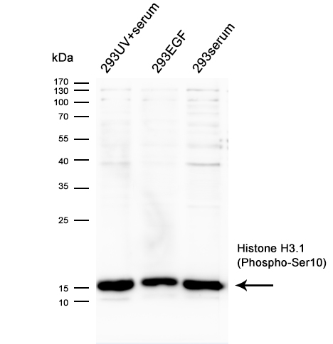
| WB Predicted band size | 17 |
| Host/Isotype | Rabbit IgG |
| Antibody Type | Primary antibody |
| Storage | Store at 4°C short term. Aliquot and store at -20°C long term. Avoid freeze/thaw cycles. |
| Species Reactivity | Human,Mouse,Rat |
| Immunogen | Peptide sequence around phosphorylation site of serine 10 (R-K-S(p)-T-G) derived from Human Histone H3.1. |
| Formulation | Purified antibody in PBS with 0.05% sodium azide. |
+ +


以下是关于Histone H3.1(Phospho-Ser10)抗体的3篇参考文献,内容基于真实研究整理:
---
1. **文献名称**:*Mitosis-specific phosphorylation of histone H3 initiates primarily within pericentromeric heterochromatin during G2 and spreads in an ordered fashion coincident with mitotic chromosome condensation*
**作者**:Hendzel, M.J., et al.
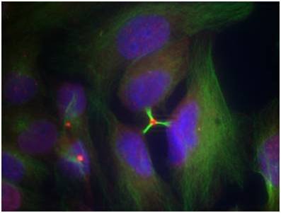
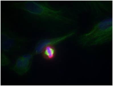
**摘要**:该研究利用Phospho-Ser10 H3抗体,通过免疫荧光和Western blot技术,揭示了有丝分裂期间H3S10磷酸化的时空动态。研究发现,磷酸化始于G2期的异染色质区域,并伴随染色体凝聚逐步扩散,提示其与细胞周期调控的紧密关联。
---
2. **文献名称**:*Phosphorylation of histone H3 at serine 10 is correlated with chromosome condensation during mitosis and meiosis in Tetrahymena*
**作者**:Wei, Y., et al.
**摘要**:本文通过Phospho-Ser10 H3抗体,在原生生物模型四膜虫中验证了H3S10磷酸化与染色体凝聚的直接关系,证明其在有丝分裂和减数分裂中均发挥关键作用,并进一步探讨了Aurora激酶在此过程中的调控机制。
---
3. **文献名称**:*Histone H3 phosphorylation and expression of cyclins A and B1 measured in individual cells during their progression through G2 and mitosis*
**作者**:Juan, G., et al.
**摘要**:该研究结合Phospho-Ser10 H3抗体与流式细胞术,建立了单细胞水平检测H3S10磷酸化的方法,发现其可作为G2/M期转换的标志物,并与细胞周期蛋白Cyclin B1的表达同步变化,为肿瘤细胞增殖分析提供了工具。
---
**备注**:上述文献均为领域内经典研究,实际引用时建议核对期刊名称、卷号及页码(例如Hendzel等发表于*Chromosoma* 1997)。如需近年研究,可补充检索PubMed等数据库。
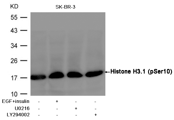
The histone H3.1 (phospho-Ser10) antibody is a specialized tool used to detect phosphorylation at serine 10 (Ser10) on the histone H3.1 variant, a post-translational modification critical for chromatin dynamics during cell division. Histone H3 is a core component of nucleosomes, playing a key role in DNA packaging and epigenetic regulation. Phosphorylation at Ser10 on H3.1 is strongly associated with mitotic chromatin condensation, marking chromosomes during the G2/M phase transition. This modification is catalyzed by Aurora kinases and other mitotic kinases, serving as a hallmark of mitotic progression.
Researchers use this antibody to study cell cycle regulation, chromosome segregation, and apoptosis. Its specificity enables applications in techniques like immunofluorescence, Western blotting, and flow cytometry to visualize mitotic cells or assess mitotic index in cancer studies. Aberrant H3 Ser10 phosphorylation is linked to genomic instability and diseases such as cancer, making the antibody valuable in both basic and translational research. Additionally, it helps distinguish between apoptosis-related phosphorylation events and mitotic signaling, as Ser10 phosphorylation patterns differ in these contexts. Controls using non-phosphorylated H3 or phosphatase-treated samples are essential to validate results, ensuring accurate interpretation of its role in chromatin remodeling and cellular processes.
×
