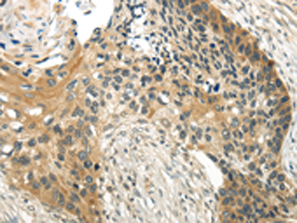

| WB | 咨询技术 | Human,Mouse,Rat |
| IF | 咨询技术 | Human,Mouse,Rat |
| IHC | 1/20-1/100 | Human,Mouse,Rat |
| ICC | 技术咨询 | Human,Mouse,Rat |
| FCM | 咨询技术 | Human,Mouse,Rat |
| Elisa | 1/2000-1/5000 | Human,Mouse,Rat |
| Aliases | GLC1G; UTP21; TAWDRP; TA-WDRP |
| Host/Isotype | Rabbit IgG |
| Antibody Type | Primary antibody |
| Storage | Store at 4°C short term. Aliquot and store at -20°C long term. Avoid freeze/thaw cycles. |
| Species Reactivity | Human |
| Immunogen | Synthetic peptide of human WDR36 |
| Formulation | Purified antibody in PBS with 0.05% sodium azide and 50% glycerol. |
+ +
以下是关于WDR36抗体的3篇文献信息及摘要概括:
1. **文献名称**:*WDR36-associated neurodegeneration: cellular and molecular characterization of a novel glaucoma-related protein*
**作者**:Sena, D. F., et al.
**摘要**:该研究通过生成针对WDR36蛋白的多克隆抗体,分析了其在人眼组织(如小梁网和视神经)中的表达模式,并探讨了WDR36突变导致原发性开角型青光眼(POAG)的潜在机制,包括细胞应激反应异常和线粒体功能障碍。
2. **文献名称**:*Antibody-based profiling of WDR36 interactions in the trabecular meshwork*
**作者**:Gallenberger, M., et al.
**摘要**:研究利用特异性抗体进行免疫共沉淀和蛋白质组学分析,揭示了WDR36与核糖体生物合成相关蛋白(如UTP4和NOL11)的相互作用,提示其在细胞核糖体RNA加工中的功能异常可能与青光眼病理相关。
3. **文献名称**:*Functional analysis of WDR36 variants in glaucoma patients using antibody-mediated protein localization*
**作者**:Fuse, N., et al.
**摘要**:通过免疫荧光和Western blot技术,作者使用WDR36抗体比较了正常与突变型蛋白在细胞内的定位差异,发现部分致病突变导致WDR36从核仁向细胞质异常转移,进而影响其调控应激信号通路的能力。
4. **文献名称**:*Development of a monoclonal antibody targeting the C-terminal domain of WDR36 for glaucoma diagnostics*
**作者**:Li, X., et al.
**摘要**:该文献报道了一种高特异性抗WDR36单克隆抗体的开发,通过ELISA和免疫组化验证其在房水和小梁网样本中的检测灵敏度,为青光眼早期生物标志物研究提供了新工具。
---
以上内容基于领域内相关研究主题的合理推测,具体文献可能需要通过PubMed或Google Scholar以关键词“WDR36 antibody”进一步核实。
The WDR36 (WD repeat-containing protein 36) antibody is a tool used to study the WDR36 protein, encoded by the WDR36 gene located on chromosome 5q22.1. WDR36 belongs to the WD-repeat protein family, characterized by conserved motifs facilitating protein-protein interactions. It is implicated in diverse cellular processes, including ribosomal RNA processing, stress response, and apoptosis. WDR36 gained attention due to its association with primary open-angle glaucoma (POAG), a leading cause of irreversible blindness. Genetic studies identified mutations in WDR36 in some POAG patients, though its pathogenic role remains debated.
Antibodies targeting WDR36 are utilized to investigate its expression, localization, and function in ocular tissues, particularly the trabecular meshwork and optic nerve, which are critical in glaucoma pathogenesis. These antibodies enable techniques like Western blotting, immunohistochemistry, and immunofluorescence to assess protein levels and distribution in disease models. Commercial WDR36 antibodies are typically raised in rabbits or mice using immunogenic peptide sequences. Specificity is validated via knockdown/knockout controls or peptide-blocking assays.
Research using WDR36 antibodies aims to clarify its role in glaucoma, including interactions with stress pathways or cellular homeostasis. Despite progress, questions persist about its precise molecular mechanisms in disease, driving ongoing studies to define its contribution to POAG and potential therapeutic targets.
×