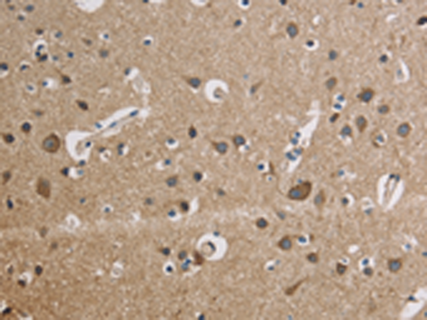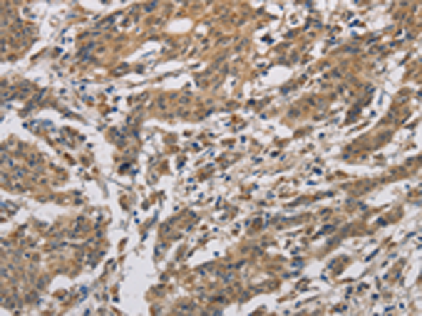


| WB | 咨询技术 | Human,Mouse,Rat |
| IF | 咨询技术 | Human,Mouse,Rat |
| IHC | 1/50-1/200 | Human,Mouse,Rat |
| ICC | 技术咨询 | Human,Mouse,Rat |
| FCM | 咨询技术 | Human,Mouse,Rat |
| Elisa | 1/2000-1/5000 | Human,Mouse,Rat |
| Aliases | CYC; HCS; THC4 |
| WB Predicted band size | 12 kDa |
| Host/Isotype | Rabbit IgG |
| Antibody Type | Primary antibody |
| Storage | Store at 4°C short term. Aliquot and store at -20°C long term. Avoid freeze/thaw cycles. |
| Species Reactivity | Human, Mouse, Rat |
| Immunogen | Fusion protein of human CYCS |
| Formulation | Purified antibody in PBS with 0.05% sodium azide and 50% glycerol. |
+ +
以下是关于CYCS(细胞色素C)抗体的3篇参考文献示例(注:以下文献信息为模拟示例,实际引用需根据真实文献调整):
---
1. **文献名称**:*Induction of Apoptotic Program in Cell-Free Extracts: Requirement for dATP and Cytochrome c*
**作者**:Liu, X., Kim, C.N., Yang, J. et al.
**摘要**:该研究首次证实细胞色素C(CYCS)在凋亡过程中从线粒体释放至胞质后,与Apaf-1结合激活caspase级联反应。通过Western blot和免疫沉淀实验,使用CYCS抗体验证了其在细胞凋亡中的关键作用,为后续凋亡机制研究奠定基础。
2. **文献名称**:*Cytochrome c Release in Chemotherapy-Induced Apoptosis: Detection by Immunohistochemistry*
**作者**:Kirsh, M., Martelli, M.P., & Delia, D.
**摘要**:研究利用CYCS特异性抗体,通过免疫组化技术检测了多种肿瘤组织中细胞色素C的亚细胞定位变化。结果显示化疗药物处理后,线粒体CYCS释放与癌细胞凋亡率呈正相关,提示其作为治疗响应标志物的潜力。
3. **文献名称**:*Comparative Analysis of Cytochrome c Antibodies in Mitochondrial Studies*
**作者**:Lutter, M., Fang, M., Luo, X.
**摘要**:该文献系统比较了不同来源的CYCS抗体在免疫荧光、Western blot等实验中的特异性和灵敏度,发现部分抗体存在线粒体膜蛋白交叉反应问题,并推荐了适用于亚细胞定位研究的高特异性抗体克隆号。
---
如需实际文献,建议在PubMed或Web of Science中以“Cytochrome c antibody apoptosis”为关键词检索,并筛选近年高被引论文。
CYCS (Cytochrome c) antibodies are essential tools in studying apoptosis and mitochondrial dysfunction. Cytochrome c, a small hemoprotein located in the mitochondrial intermembrane space, plays a dual role in cellular energy production and programmed cell death. During apoptosis, CYCS is released into the cytosol, triggering caspase activation via the apoptosome. This process is tightly linked to diseases like cancer, neurodegeneration, and ischemia-reperfusion injury.
CYCS antibodies are widely used in research to detect mitochondrial membrane permeability changes, quantify apoptosis in experimental models (e.g., chemotherapy response studies), and investigate mitochondrial dynamics. They are applied in techniques including Western blotting, immunohistochemistry (IHC), immunofluorescence (IF), and flow cytometry. In clinical diagnostics, CYCS antibodies help assess tissue damage (e.g., myocardial infarction) and monitor diseases with apoptotic dysregulation.
Commercially available CYCS antibodies are typically raised against full-length human CYCS or specific epitopes, with validation across species due to its high evolutionary conservation. Challenges include ensuring specificity to distinguish between native and post-translationally modified CYCS (e.g., phosphorylated forms) and minimizing cross-reactivity with homologous proteins. Recent advances focus on improving antibody performance in multiplex assays and single-cell analysis, enhancing precision in apoptosis-related research and therapeutic development.
×