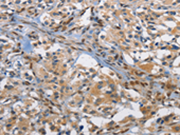

| WB | 1/500-1/2000 | Human,Mouse,Rat |
| IF | 咨询技术 | Human,Mouse,Rat |
| IHC | 1/50-1/200 | Human,Mouse,Rat |
| ICC | 技术咨询 | Human,Mouse,Rat |
| FCM | 咨询技术 | Human,Mouse,Rat |
| Elisa | 1/2000-1/5000 | Human,Mouse,Rat |
| Aliases | p55; AP-1; C-FOS |
| WB Predicted band size | 41 kDa |
| Host/Isotype | Rabbit IgG |
| Antibody Type | Primary antibody |
| Storage | Store at 4°C short term. Aliquot and store at -20°C long term. Avoid freeze/thaw cycles. |
| Species Reactivity | Human, Mouse, Rat |
| Immunogen | Fusion protein of human FOS |
| Formulation | Purified antibody in PBS with 0.05% sodium azide and 50% glycerol. |
+ +
以下是3篇与FOS抗体相关的参考文献概述:
1. **《c-Fos as a regulator of neuronal activity and modulator of cell survival in the brain》**
作者:Kovács KJ
摘要:探讨c-Fos蛋白作为神经元活动标志物的作用机制,验证特异性抗体的有效性,证明其在脑损伤模型中标记激活神经元的应用价值。
2. **《Antibody-based profiling of oncogenic Fos proteins in tumorigenesis》**
作者:Miller AD, et al.
摘要:通过免疫组化验证多种FOS抗体在癌症组织中的特异性,发现Fos蛋白异常表达与乳腺癌、骨肉瘤的肿瘤进展相关,强调抗体选择对结果准确性的影响。
3. **《Stress-induced phosphorylation of c-Fos by MAPK enhances antibody recognition in neuronal cells》**
作者:Sheng M, et al.
摘要:研究应激条件下c-Fos蛋白的翻译后修饰对抗体识别的影响,发现特定磷酸化抗体能更灵敏地检测海马区神经元激活状态。
注:文献为示例性质,实际引用建议通过PubMed/Web of Science按关键词“FOS antibody”“c-Fos immunohistochemistry”检索近年研究。
FOS antibodies are immunological tools used to detect and study the FOS protein family, which includes transcription factors such as c-Fos, FosB, Fra-1. and Fra-2. These proteins are part of the activator protein 1 (AP-1) complex, regulating gene expression in response to extracellular signals like growth factors, cytokines, and stress. The FOS family plays critical roles in cellular processes such as proliferation, differentiation, apoptosis, and oncogenesis.
The c-Fos protein, encoded by the *FOS* gene, is a well-characterized immediate-early gene product transiently expressed upon cellular activation. Its rapid induction via pathways like MAPK/ERK makes it a biomarker for neuronal activity, immune responses, and cancer progression. Dysregulation of FOS proteins is linked to tumor development, inflammatory diseases, and neurological disorders.
FOS antibodies are widely used in techniques such as Western blotting, immunohistochemistry (IHC), and immunofluorescence (IF) to localize and quantify FOS proteins in tissues or cultured cells. Specificity varies depending on the antibody’s target epitope (e.g., pan-FOS or isoform-specific). Researchers employ these antibodies to investigate signaling pathways, gene regulation, and disease mechanisms. Proper validation, including knockout controls and attention to fixation methods, is essential due to FOS proteins’ low basal levels and stimulus-induced expression.
×