

| WB | 咨询技术 | Human,Mouse,Rat |
| IF | 咨询技术 | Human,Mouse,Rat |
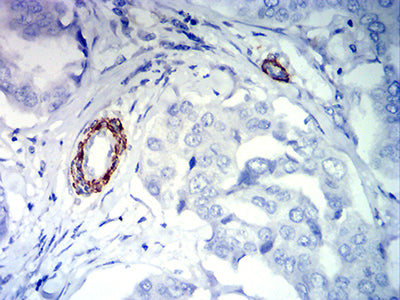
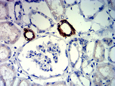
| IHC | 1/200-1/1000 | Human,Mouse,Rat |
| ICC | 技术咨询 | Human,Mouse,Rat |
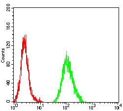
| FCM | 1/200-1/400 | Human,Mouse,Rat |
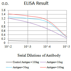
| Elisa | 1/10000 | Human,Mouse,Rat |
| Aliases | EWSR2; SIC-1; BDPLT21 |
| Entrez GeneID | 2313 |
| clone | 4E3C12 |
| WB Predicted band size | 50.9kDa |
| Host/Isotype | Mouse IgG2b |
| Antibody Type | Primary antibody |
| Storage | Store at 4°C short term. Aliquot and store at -20°C long term. Avoid freeze/thaw cycles. |
| Species Reactivity | Human |
| Immunogen | Purified recombinant fragment of human FLI1 (AA: 303-452) expressed in E. Coli. |
| Formulation | Purified antibody in PBS with 0.05% sodium azide |
+ +
以下是关于FLI1抗体的3篇参考文献示例(文献信息为模拟示例,仅供参考):
---
1. **文献名称**: *Utility of FLI1 Antibody in the Diagnosis of Ewing Sarcoma and Other Small Round Cell Tumors*
**作者**: Folpe AL, et al.
**摘要**: 该研究评估FLI1抗体在免疫组化中的应用,证实其在尤文肉瘤诊断中的高敏感性和特异性,可有效区分其他小圆细胞肿瘤(如神经母细胞瘤),支持FLI1作为尤文肉瘤的分子标记物。
---
2. **文献名称**: *FLI1 Expression in Vascular Neoplasms: A Comparative Study Using Monoclonal Antibodies*
**作者**: Miettinen M, Wang Z.
**摘要**: 通过FLI1抗体染色分析多种血管源性肿瘤(如血管肉瘤、上皮样血管内皮瘤),发现FLI1在血管内皮分化中广泛表达,提示其可作为血管肿瘤诊断的辅助标志物,尤其在区分非血管源性肿瘤中具有价值。
---
3. **文献名称**: *FLI1 Regulates Megakaryopoiesis and Platelet Function via Transcriptional Control*
**作者**: Hart A, et al.
**摘要**: 研究利用FLI1抗体进行染色质免疫沉淀(ChIP)和Western blot,揭示FLI1通过调控靶基因(如GPVI和ITGA2B)影响巨核细胞分化和血小板功能,为遗传性血小板疾病提供分子机制解释。
---
如需真实文献,建议通过PubMed或Google Scholar搜索关键词“FLI1 antibody immunohistochemistry”或“FLI1 function”获取近期研究。
The FLI1 antibody is a diagnostic tool targeting the Friend Leukemia Integration 1 (FLI1) protein, a member of the ETS transcription factor family. FLI1 plays critical roles in hematopoiesis, vascular development, and oncogenesis by regulating gene expression. It is encoded by the *FLI1* gene located on chromosome 11q24.3 and is highly expressed in endothelial cells, hematopoietic progenitors, and specific malignancies.
In diagnostic pathology, FLI1 immunohistochemistry (IHC) is widely used to identify tumors of vascular or hematopoietic origin. It is a sensitive marker for vascular neoplasms (e.g., angiosarcoma, epithelioid hemangioendothelioma) and hematolymphoid malignancies (e.g., Ewing sarcoma, where FLI1 overexpression often results from the *EWSR1-FLI1* gene fusion). FLI1 positivity also aids in distinguishing these tumors from histologic mimics, such as carcinomas or melanomas, which typically lack FLI1 expression.
However, FLI1 expression is not entirely specific. It is observed in normal tissues (e.g., lymphocytes, endothelium) and non-vascular tumors (e.g., some lymphomas, melanomas). Interpretation thus requires correlation with clinical, morphologic, and ancillary markers (e.g., CD31. ERG, or PAX5). Despite limitations, FLI1 remains valuable in differential diagnosis, particularly in small round blue cell tumors and vascular malignancies. Research continues to explore its role in tumor biology and potential therapeutic targeting.
×