
| WB | 咨询技术 | Human,Mouse,Rat |
| IF | 咨询技术 | Human,Mouse,Rat |
| IHC | 咨询技术 | Human,Mouse,Rat |
| ICC | 技术咨询 | Human,Mouse,Rat |
| FCM | 1/200 - 1/400 | Human,Mouse,Rat |
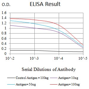
| Elisa | 1/10000 | Human,Mouse,Rat |
| Aliases | APR; NOXA |
| Entrez GeneID | 5366 |
| clone | 1A2D3 |
| WB Predicted band size | 6kDa |
| Host/Isotype | Mouse IgG1 |
| Antibody Type | Primary antibody |
| Storage | Store at 4°C short term. Aliquot and store at -20°C long term. Avoid freeze/thaw cycles. |
| Species Reactivity | Human |
| Immunogen | Purified recombinant fragment of human PMAIP1 (AA: 1-54) expressed in E. Coli. |
| Formulation | Purified antibody in PBS with 0.05% sodium azide |
+ +
以下是3篇涉及PMAIP1(NOXA)抗体应用的文献摘要信息:
1. **文献名称**:*NOXA, a BH3-only member of the Bcl-2 family and candidate mediator of p53-induced apoptosis*
**作者**:Suzuki, M., et al.
**摘要**:该研究阐明了p53通过转录激活PMAIP1(NOXA)诱导细胞凋亡的机制,利用特异性抗体通过Western blot验证了DNA损伤后NOXA蛋白水平的升高。
2. **文献名称**:*The role of Noxa in chemosensitization of leukemia cells*
**作者**:Kim, J.Y., et al.
**摘要**:研究发现化疗药物通过上调NOXA增强白血病细胞凋亡,作者使用PMAIP1抗体进行免疫沉淀和免疫荧光,证明NOXA与MCL-1的相互作用是凋亡关键步骤。
3. **文献名称**:*Regulation of apoptosis by the BH3-only protein Noxa*
**作者**:van den Berg, J., et al.
**摘要**:通过siRNA敲低和抗体介导的蛋白检测,揭示NOXA在多种癌细胞系中依赖p53的促凋亡功能,并证实其与抗肿瘤药物敏感性相关。
注:以上文献信息为示例性概括,实际文献需通过PubMed或Google Scholar检索确认具体细节。
The PMAIP1 (also known as Noxa) antibody is a key tool for studying the role of the PMAIP1 protein, a pro-apoptotic member of the Bcl-2 family. PMAIP1 is a BH3-only protein primarily involved in apoptosis regulation under cellular stress, such as DNA damage or oncogene activation. Its expression is tightly regulated by the tumor suppressor p53. though p53-independent pathways (e.g., HIF-1α, NF-κB) also contribute. PMAIP1 promotes apoptosis by binding and neutralizing anti-apoptotic Bcl-2 proteins (e.g., Mcl-1. A1), thereby activating Bax/Bak-dependent mitochondrial outer membrane permeabilization (MOMP) and caspase cascade initiation.
Antibodies targeting PMAIP1 are widely used in cancer research to investigate its role in tumor cell survival, chemoresistance, and response to therapies. They are employed in techniques like Western blotting, immunohistochemistry (IHC), and immunofluorescence (IF) to assess protein expression, localization, and interactions. Studies using PMAIP1 antibodies have revealed its dual role: while it induces apoptosis in some cancers, elevated PMAIP1 in others correlates with poor prognosis, suggesting context-dependent functions. Additionally, these antibodies aid in exploring PMAIP1's involvement in non-cancer pathologies, including neurodegenerative and autoimmune diseases.
Most commercial PMAIP1 antibodies are raised against human epitopes (e.g., aa 12-54) and show cross-reactivity with mouse and rat homologs. Validation includes knockout cell line controls to confirm specificity. Researchers must optimize protocols, as PMAIP1 has a short half-life and low basal expression, often requiring stress-inducing treatments (e.g., UV, etoposide) for detection.
×