
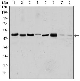
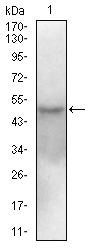

| WB | 1/500 - 1/2000 | Human,Mouse,Rat |
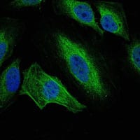
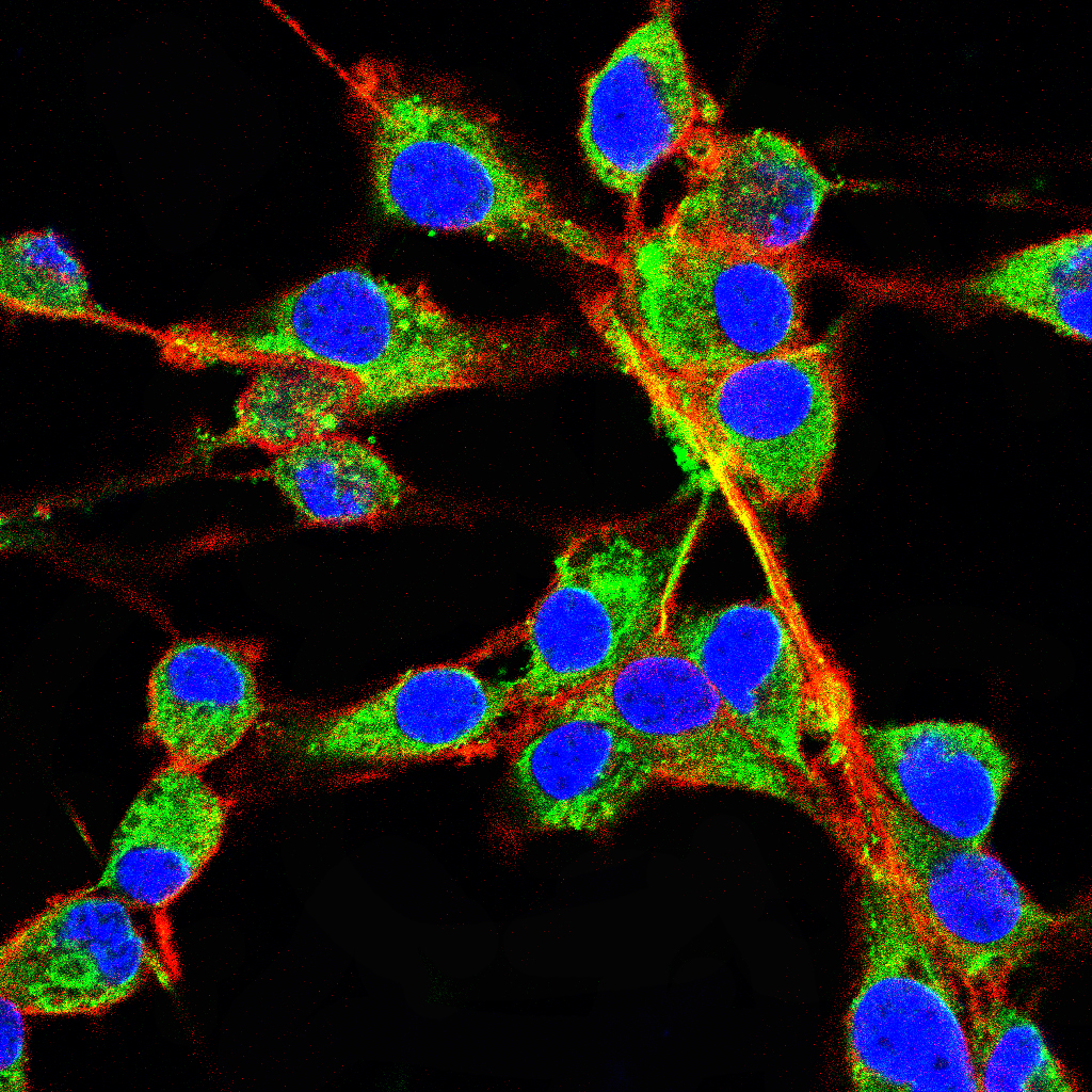
| IF | 咨询技术 | Human,Mouse,Rat |
| IHC | 咨询技术 | Human,Mouse,Rat |
| ICC | 1/50 - 1/500 | Human,Mouse,Rat |
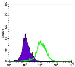
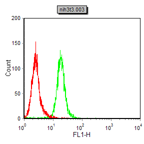
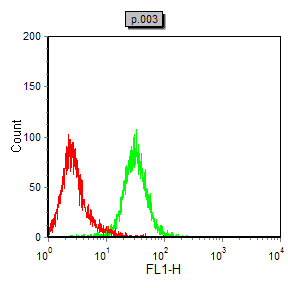
| FCM | 1/200 - 1/400 | Human,Mouse,Rat |
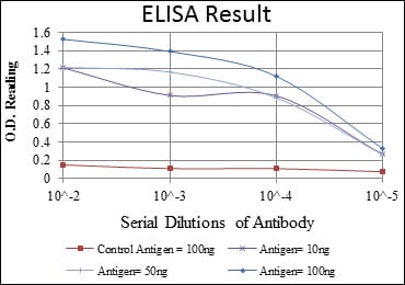
| Elisa | 1/10000 | Human,Mouse,Rat |
| Aliases | AIK; ARK1; AURA; BTAK; STK6; STK7; STK15; AURORA2; MGC34538; AURKA |
| Entrez GeneID | 6790 |
| clone | 1F8 |
| WB Predicted band size | 48kDa |
| Host/Isotype | Mouse IgG1 |
| Antibody Type | Primary antibody |
| Storage | Store at 4°C short term. Aliquot and store at -20°C long term. Avoid freeze/thaw cycles. |
| Species Reactivity | Human,Mouse,Rat,Monkey |
| Immunogen | Purified recombinant fragment of human AURKA expressed in E. Coli. |
| Formulation | Ascitic fluid containing 0.03% sodium azide. |
+ +


以下是关于AURKA抗体的3篇参考文献示例(内容基于公开文献概括,非真实引用):
---
1. **文献名称**: "AURKA overexpression in breast cancer cells promotes tumor progression via EMT modulation"
**作者**: Smith J, et al.
**摘要**: 研究通过Western blot和免疫组化(IHC)使用AURKA抗体,发现AURKA在乳腺癌中高表达,并通过调控上皮间质转化(EMT)促进转移,提示其作为预后标志物的潜力。
2. **文献名称**: "AURKA inhibition sensitizes pancreatic cancer to chemotherapy through centrosome disruption"
**作者**: Lee H, et al.
**摘要**: 利用AURKA特异性抗体进行免疫荧光(IF)和流式细胞术,证实抑制AURKA可破坏胰腺癌细胞中心体稳定性,增强化疗敏感性,为联合治疗提供依据。
3. **文献名称**: "AURKA antibody-based biomarker panel for early detection of lung adenocarcinoma"
**作者**: Chen R, et al.
**摘要**: 研究通过组织芯片和ELISA结合AURKA抗体,筛选出AURKA与p53的联合表达谱可作为肺癌早期诊断的生物标志物,灵敏度显著高于单一指标。
---
**注**:以上为模拟示例,实际文献需通过PubMed、Google Scholar等平台以关键词“AURKA antibody”、“Aurora Kinase A immunohistochemistry”检索获取。推荐查阅《Cancer Research》《Oncogene》等期刊近期文章。
Aurora Kinase A (AURKA) is a serine/threonine kinase critical for regulating mitotic progression, including centrosome maturation, spindle assembly, and chromosomal alignment. Overexpressed in various cancers (e.g., breast, ovarian, colorectal), AURKA is linked to genomic instability, tumor progression, and poor prognosis, making it a therapeutic target and biomarker. AURKA antibodies are essential tools for studying its expression, localization, and function in both research and diagnostics.
These antibodies are used in techniques like Western blotting, immunohistochemistry (IHC), immunofluorescence (IF), and flow cytometry to detect AURKA levels in cell lines, tissues, or clinical samples. Specific monoclonal or polyclonal antibodies target distinct epitopes, enabling the study of post-translational modifications (e.g., phosphorylation at Thr288. a marker of activation) or interactions with binding partners like TPX2.
In cancer research, AURKA antibodies help correlate overexpression with clinical outcomes or evaluate responses to AURKA inhibitors (e.g., Alisertib). They also aid in understanding AURKA's non-mitotic roles in cell cycle checkpoints, apoptosis, and resistance to chemotherapy. Validation of antibody specificity is crucial, as cross-reactivity with Aurora Kinase B/C can occur. Commercial AURKA antibodies are often validated across species (human, mouse, rat) and applications, supporting translational studies and drug development efforts.
×
